- FREELY available online or in PDF format. Print books can be purchased here.
- All royalties from book sales go to Cancer Research UK. Want to read for free but still donate to Cancer Research UK? Click here.
-

Chapter 4: Country Life — Repair and Replication
For the last quarter of a century, the Clare Hall Laboratories have been a highly productive focal point for research into DNA repair and replication. Never numbering more than 10 small groups, scientists at Clare Hall look out over a landscape of fields, farms, and riding stables, shortly after the last suburbs of North London give way to the placid Hertfordshire countryside. The muddy tranquillity of their surroundings is only slightly disturbed by the distant hum of traffic from the M25 and the A1 motorways, which pass nearby. It is a far cry from the urban buzz surrounding Clare Hall’s metropolitan big brother in Lincoln’s Inn Fields.
By rights, Clare Hall should not have been a success. It was isolated, too small, too focussed—any of these factors could have been a death sentence. Although the importance of the steady financial support of the ICRF and, latterly, Cancer Research UK, should not be underestimated, the many achievements of those working there are a testament to motivated, smart science, fierce collegial loyalty, and a dedicated and exacting Director.
Unfortunately, a chapter extolling the talents and virtues of all of the inhabitants of Clare Hall would be impossibly long and most likely very tedious; a litany of success can be as good a soporific as any other list. The four protagonists in the pages that follow have therefore been selected for two reasons. Firstly, the two men responsible for the birth and development of Clare Hall were both in the vanguard of the DNA revolution, and their work marches with the evolution of research into replication and repair. Secondly, the stories that complete the chapter are perfect illustrations of what Clare Hall does best: hiring very good people, and then trusting their abilities to turn ambitious, long-term projects from ugly ducklings into swans.
John Cairns and Mill Hill, ‘That Awful Place’
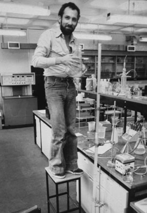
Clare Hall rose from the ashes of the ICRF Mill Hill Laboratory, a suburban outpost situated in North London at the far end of the Northern Line. Hunkered down on the doorstep of the giant MRC National Institute for Medical Research, the Mill Hill laboratories had been rented from the MRC on a 50-year lease in 1936, and for a while had been quite successful. However, like the rest of the ICRF, by the time of Michael Stoker’s appointment as Director in 1969, the labs had fallen into a sleepy complacency, enlivened only by the daily ritual of Afternoon Tea in the library, complete with crustless cucumber sandwiches, and the arrival and departure of the unit Director in his Bristol 405, a sleekly beautiful four-door sports saloon that was its owner’s pride and joy. The lab was rudely awakened from its genteel slumbering in 1973 by the arrival of a molecular biologist eager to turn his considerable talents to cancer research. John Cairns, the ex-Director of the Cold Spring Harbor lab, was about to shake things up rather spectacularly, in the process laying down the foundations on which Clare Hall’s future successes were built.
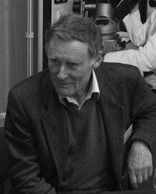
John Cairns, 2002.
(Photograph courtesy of CRUK London Research Institute Archives.)
John Cairns is a fascinating figure in the landscape of 20th century biology. Switching fields with the ease of a small boy scrambling over a fence, he left his mark on everything to which he turned his hand, leaving a trail of significant papers in each discipline before moving on to the next. As an added bonus, he is one of the most thoughtful and elegant chroniclers of the molecular biology revolution, in which he was both a distinguished participant and an observer, and his subsequent writings on the biology and epidemiology of cancer are still relevant and thought-provoking today. The esteem and affection in which he and his wife Elfie are held by their many friends and acquaintances are a tribute to their unique combination of charm, intelligence, and hospitality.
The grandson of a Master of Balliol, son of the hugely talented and influential brain surgeon Sir Hugh Cairns, godson of Sir Charles Sherrington (Nobel Laureate and founder of neurobiology), John was born into the intellectual aristocracy of Britain and had the aptitude and connections to have followed his father into a stellar career as a surgeon. Instead, after qualifying as a doctor, he turned his back on silver spoonery, and by the mid-1950s was a successful academic virologist at the newly established University of Canberra.
In 1957, John left Elfie and his young family in Canberra for a four-month sabbatical at Caltech, and by a combination of good luck and poverty, found himself at the epicentre of the DNA revolution. Unable to afford the exorbitant room rates at the Caltech Faculty Club, John was rescued from homelessness by Jan Drake, one of Renato Dulbecco’s PhD students, and as a result found himself eating dinner most nights in an apartment shared by Drake, Matt Meselson, and Howard Temin during the period in which Meselson and Frank Stahl were doing what Cairns subsequently christened “the most beautiful experiment in biology.”
Meselson and Stahl’s 1958 paper, “The Replication of DNA in Escherichia coli,” was the first big step towards the wider acceptance that DNA really was the hereditary material of the cell, and not just a boring structural component of chromosomes, as many of the biochemists and physical chemists who worked on it had previously decided. Oswald Avery, Colin MacLeod, and Maclyn McCarty’s 1944 experiment showing that the “transforming principle” of Streptococcus pneumoniae was DNA, and Al Hershey and Martha Chase’s more recent demonstration in 1952 that the infectious material of a phage was also DNA, had not made much of an impact outside the world of molecular biology, perhaps being regarded with suspicion as new-fangled genetic sleights of hand. Watson and Crick had suggested in 1953 that the invariant base pairing of the rungs of the DNA ladder, with cytosine (C) always with guanine (G), and adenine (A) always with thymine (T), provided a great mechanism by which one strand could produce a complementary copy of itself, but it was just another theory until Meselson and Stahl gave it reality. (It should be noted that the invariant C:G and A:T ratio between the bases was originally observed by Erwin Chargaff, but he failed to realise the significance. He was, consequently, very grumpy about DNA and Watson and Crick for much of the rest of his life.) Although they were careful not to overinterpret their experiment, referring cautiously to two DNA subunits, Meselson and Stahl’s work showed that the two strands of the DNA double helix came apart and were each used as a template to make a new strand. The result was a duplication of the original molecule, with each duplicate containing one old and one newly synthesised strand; in other words, DNA was replicated semi-conservatively.
John Cairns had a wonderful time at Caltech. He got to grips with molecular biology whilst doing a lot of washing up at the Meselson ménage, attended the phage course (he is remembered by one of the other participants as “admirably modest, generous and collegial, but […] it was rather as if Sir Lancelot had decided to attend the school prom”) (Susman 1995), and survived an attempt at manslaughter by camping, Delbrück style (“as I waited through the night for the dawn to come, I thought I would freeze to death”) (Cairns et al. 2007). It was not entirely surprising that upon his return to Canberra, he was an addicted molecular biologist and switched the focus of his science accordingly. Following another, longer sabbatical with Al Hershey at Cold Spring Harbor in 1960, John published the definitive proof that DNA replicated as a single double helix and that replication in bacteria proceeded bidirectionally round the circular E. coli chromosome from a single origin. In the process, he invented the technique of DNA autoradiography, visualising DNA by radioactively labeling it with tritiated thymidine. His autoradiographs of E. coli DNA caught in the act of replication are simultaneously a technical tour de force and a monument to extreme patience and persistence. John himself dismissed this minor classic as “an act of tidying (rather like washing-up, in fact)” (Cairns 1980).
In 1963, John moved his family permanently to Cold Spring Harbor because he’d been inveigled into becoming the laboratory’s Director, little realising that the place was on its knees both financially and structurally and that he had been hired to oversee its downfall. As Matt Meselson recalls: “I think John was expecting the same gentlemanly standards that John himself would have practised; instead he walked into a nightmare. But he did get the place out of the red, which was an enormous accomplishment. It was about to expire. Not only expire, but to expire in sewage” (Meselson, CSH Oral History). After five years of “the most hideous time,” during which he greatly annoyed the laboratory’s detractors by getting the place back on its feet again, John handed over the Directorship to Jim Watson and got back to what he loved best, doing experiments.
The project that John began impinged on a parallel, biochemical strand in the story of DNA—the question of how the molecule was made. Although Meselson and Stahl had shown that semi-conservative replication could occur, the nuts and bolts of exactly how DNA replicated itself was a very biochemical matter. Where there is a process, there has to be an enzyme to perform it, and fittingly, the eureka enzyme of DNA replication, DNA polymerase, was discovered by one of the greatest biochemists of the 20th century, Arthur Kornberg.
Kornberg, as the host of stories lovingly propagated by his friends and colleagues testifies, was in his own way as charismatic as Delbrück but was an implacable dragon in the laboratory. His allegiance to biochemistry meant that anything other than total commitment and experimental rigour was unacceptable, encapsulated in the maxim that he adopted from his fellow biochemist Efraim Racker: “Don’t waste clean thinking on dirty enzymes”; and the first of his famous Ten Commandments of Enzymology: “Thou shalt rely on enzymology to resolve and reconstitute biologic events.” Kornberg’s genius was to realise that everything, even the most complex molecular machine, could be purified out of the cell, and that reconstituting such machines in the relative tranquillity of the test tube was the way to understand how they worked. His interest in DNA arose because he wanted to know how nucleotides, which he and others had shown were the building blocks of DNA chains, were put together. The fact that DNA might be the hereditary material and that Watson and Crick had proposed a model for its double-helical structure was interesting, but the possibility of proving that DNA synthesis was not one of life’s untouchable mysteries, but as much an enzymatic process as the fermentation of yeast, was what initially drove him. From 1954, when he was appointed Chair of the Microbiology Department at Washington University in St Louis, Kornberg worked towards detecting the polymerisation of nucleotides, beginning first with RNA and then widening his studies to include DNA synthesis.
After some years of labour, Kornberg and his lab got a hint that they were on the right track when they managed to get an E. coli cellular extract to synthesise a vanishingly small amount of polymerised DNA. This was all Kornberg needed. Two years later, when he won the 1959 Nobel Prize along with his mentor Severo Ochoa, “for their discovery of the mechanisms in the biological synthesis of ribonucleic acid and deoxyribonucleic acid,” he recalled that “through this tiny crack we tried to drive a wedge, and the hammer was enzyme purification” (Kornberg 1960). The enzyme his lab purified, DNA polymerase, was able to stitch together nucleotides to make a new DNA strand, but crucially, only in the presence of an existing strand; the substrate, DNA, was directing its own synthesis, an event unprecedented in the annals of enzymology, but exactly as the semi-conservative replication observed by Meselson and Stahl predicted.
Up until John Cairns’s release from the burden of the Cold Spring Harbor Directorship, everybody believed that the Kornberg DNA polymerase was the solo star of the replication show, doing everything by itself. John and his technician Paula deLucia changed all this by showing that although the Kornberg DNA polymerase might be able to synthesise DNA in a test tube, it was certainly not doing the real work of replication in E. coli. In so doing, they scored a satisfying home run for molecular biology, showing to the biochemists that bacterial genetics was as important a tool as enzyme purification.
Having heard that Roy Curtis at Oak Ridge National Laboratory had isolated a mutant of E. coli that made miniature daughter cells containing no DNA but lots of Kornberg polymerase, John set Paula the difficult task of finding the reciprocal mutant of E. coli, one that lacked Kornberg’s polymerase but was perfectly viable. This she did, and the mutant, which John christened polA in homonymic tribute to Paula’s technical virtuosity, caused a flurry of biochemical activity resulting in the isolation of two further E. coli DNA polymerases, one of which, DNA polymerase III, was the real replicative enzyme. Arthur Kornberg’s chagrin that a mere molecular biologist had managed to show something so important without so much as partially purifying a protein, was no doubt mollified by the fact that both new polymerases were identified by his son Tom, whilst a graduate student in Malcolm Gefter’s lab at Columbia.
The business of bacterial DNA replication was not to occupy John for much longer. Keeping Cold Spring Harbor afloat had comprehensively emptied the coffers of the Cairns family, and in 1972, John started job-hunting back in the United Kingdom, where his children could finish their educations for rather less than in the States. In order to be on the spot, he arranged a year’s sabbatical with Michael Stoker at Lincoln’s Inn Fields. It was not long before Stoker realised that John was the perfect solution to a problem that had been dogging him for some time—whom to appoint as the new head of “that awful place” Mill Hill.
In John’s words, Mill Hill “was full of people who had decided that safety lay in not doing anything […]. I had to terrorise them, but after Cold Spring Harbor that was easy peasy!” The new laboratory Director, in addition to frightening the life out of the old staff, abolished the cucumber sandwiches and the segregated tea rooms and imported a whole slew of young, ambitious developmental biologists, whose story is related in detail in Chapter 9. John also changed the direction of his own research once again and started thinking in depth regarding cancer. The switch in emphasis gave rise to a rather good book, Cancer: Science and Society, published in 1978, and yet another distinguished career in cancer epidemiology and public health research. In the short term, however, John’s work on DNA replication in bacteria morphed into studying another aspect of the molecule’s lifestyle: how it repaired itself when damaged.
The mere existence of DNA repair had come as a big surprise to many people. In the first half of the 20th century, genes were a concept, rather than a fact, and working in such an information vacuum meant that some interesting misconceptions had arisen. One such, in part propagated by the 1935 paper by Max Delbrück, Nikolai Timoféeff-Ressovsky, and Karl Zimmer that had inspired Schrödinger’s What Is Life?, was that it was incredibly hard to mutate genes. To create any kind of change, exposure to extreme stress such as high-intensity ionising radiation was thought to be necessary, because genes were viewed as extremely stable entities, heavily protected from the outside world in order to keep their precious cargo of genetic information intact. This was backed up by the very low rate of spontaneous mutation seen in the organisms, such as the fruit fly, in which the early geneticists did most of their work.
Delbrück and his colleagues had been heavily influenced by discussions with Hermann Muller, who in 1927 had been the first to show that X rays produced high mutation rates in fruit flies. Mutation being the lifeblood of genetics, Muller’s findings had been taken up with great enthusiasm by his field as an extraordinarily powerful tool (he was eventually awarded the 1946 Nobel Prize for Physiology or Medicine). As a spin-off from Muller’s work, the study of how radiation affected living things had become a discipline in itself by the 1930s, and the field expanded hugely over the following decade, thanks to the U.S. Atomic Energy Commission, which lavishly funded radiation biology during and after the Second World War in an effort to find ways to counteract the lethal effects of the atomic bomb. However, there were problems with radiobiology. Although it was plain that ionizing radiation, and also ultraviolet light and some particularly nasty chemicals such as mustard gas, must be damaging genes, nobody had a clue how. Matters were not helped by the field itself. Innovative and thoughtful radiation biologists did exist, but the excessively generous funding had resulted in an accumulation of mediocrity, with some labs content to repeat the same experiments again and again for no reason other than that they counted as data, however useless. Radiobiology gained a reputation for existing in a state of bloated torpor, and research into the nature of genetic mutation slumbered alongside it.
What everybody had missed, and could probably not have predicted before DNA came onto the scene, was that far from being swaddled tenderly in the warm, cosy environs of the nucleus, DNA was continually buffeted by storms of passing mutagens. The reason for the low rate of spontaneous mutation was not that genes existed behind a firewall, but that they were dynamically stable. Like a circus plate-spinner, the cell keeps its genetic information intact because it continually surveys its DNA, detects any damage, and repairs it. Jumping ahead in time, David Lane and Lionel Crawford’s p53 protein is a sophisticated gatekeeper of this determination to preserve an unblemished genome.
Because sunlight and life go hand-in-hand, enzymatic photoreactivation of DNA after ultraviolet (UV) irradiation is probably the oldest repair mechanism in existence. It was also the first identified, stumbled on inadvertently by Albert Kelner at Cold Spring Harbor, and Renato Dulbecco during his time in Salvador Luria’s lab in Bloomington, Indiana. Both Kelner and Dulbecco had been greatly frustrated by their attempts to examine the survival of, respectively, bacteria and phage, following UV irradiation. Both men found that their experiments were completely irreproducible. Sometimes, irradiation resulted in perfect survival curves, where colony number was inversely correlated with the intensity of UV treatment, but sometimes, the curves were all over the place, with seemingly random numbers of survivors cropping up even after punitive UV treatment. After months of checking and rechecking his experimental protocol, Kelner in desperation moved his bacterial plates into a cold room, so that he could work out, using a series of water baths, whether temperature played any part in the process. To his surprise, all of the survival curves began to behave perfectly, but shortly afterwards, the penny dropped: The cold room was windowless, in contrast to his previous lab, and the more sunlight the plates saw, the more colonies grew on them. Dulbecco, with the same problem in his phage experiments, and knowing of Kelner’s work, came to the same conclusion, that daylight could induce repair of UV damage, and their two papers on the subject were published in 1949.
That DNA was the substrate for photoreactivation was not revealed until 1956, by Sol Goodgal and Stan Rupert at Johns Hopkins in Baltimore. The two men showed that extracts of E. coli cells, when mashed up and added to UV-irradiated DNA from the bacterium Haemophilus influenzae, were able to repair the UV damage, as long as the reaction was exposed to daylight. The inducible repair activity in the E. coli extract was clearly due to an enzyme of some kind, because it could be killed by heating, but it took until 1978 to find what the enzyme was, as it was present in such small quantities that it proved almost impossible to purify. In the meantime, however, biophysical analysis had shown that the lesion in DNA on which the mystery enzyme acted was a pyrimidine dimer, an unholy alliance formed between two neighbouring cytosine or thymine bases on the same strand of DNA. Such dimers were very common and were dangerous because they were unable to base-pair properly, leading to errors of replication, and hence mutation. Photoreactivation repaired pyrimidine dimers by resetting them back into their monomeric states, where they were once more able to recognise their partners on the opposite strand of the double helix and could be replicated correctly.
Photoreactivation was just the tip of the repair iceberg. In 1964, Dick Boyce and Paul Howard-Flanders at Yale, and Bill Carrier and Dick Setlow at Oak Ridge National Laboratory, with several other labs hot on their heels, independently discovered an alternative mechanism for ridding the cell of pyrimidine dimers: nucleotide excision repair. Rather than rescuing the bases by remonomerising them, cells could simply cut them out, together with a short stretch of the sequence in which they were embedded, leaving a gap that could be filled in using the opposite strand of the double helix as a template.
Excision repair turned out to be able to fix a lot of other lesions besides pyrimidine dimers, and its discovery prompted a new theory to explain why DNA was double-helical. As those working on the biochemistry of replication and transcription of DNA were discovering, unwinding the strands of a double helix is a difficult topological feat; simply loosening the twist in one place would result in tightening the twist in another, and dissecting the enzymatic contortions that cells have evolved to avoid hopeless tangling has kept a legion of biochemists occupied for many years. Why would a cell go to the bother of a double helix at all, when one copy of the genetic information per chromosome was surely enough? The repair field thought it had the answer. In a Scientific American article from 1967, Bob Haynes and Phil Hanawalt suggested that “redundancy is a familiar strategem to designers of error-detecting and error-correcting codes. If a portion of one strand of the DNA helix were damaged, the information in that portion could be retrieved from the complementary strand” (Hanawalt and Haynes 1967).
Asserting that the mighty double helix existed because it was the best way to correct errors in the genome was testimony to the excitement in the new field. Shedding many of the outdated concepts of radiobiology, repair enthusiasts had realised that the discovery of DNA-specific enzymes, such as Kornberg’s DNA polymerase and the DNA-cleaving nucleases, meant that the genome was as subject to enzymatic modification as any other cellular substrate, and therefore many types of enzymatically driven repair mechanisms were likely to exist. Those prepared to branch out into a bit of genetics, to find useful mutants, and biochemistry, to characterise the processes they revealed, began mining a rich seam of unknown enzymes and pathways.
In 1967, DNA repair made it into humans, when Jim Cleaver, working at the University of California, San Francisco, realised that xeroderma pigmentosum (XP), a genetic disease causing sun sensitivity and predisposition to skin cancer, was caused by defects in excision repair. The discovery provoked elation in everybody working on DNA in bacteria, from the molecular biologists right through to the biochemists, whose sentiments were encapsulated by Bob Haynes: “I can vividly remember saying to several people, thank God, we’ve now got a disease. We’ll be able to get money from the NIH because DNA repair is relevant to a human disease!” (Friedberg 1997).
Rather surprisingly, the link between DNA repair and cancer was not pursued with much enthusiasm until nearly a quarter of a century later, when, as we shall see below in this chapter, DNA repair defects were shown to be major contributors to colon and breast cancer susceptibility. Although there were some technical obstacles, the main reason appears to have been a lack of communication between fields. Theodore Boveri had proposed that cancer was a disease caused by mutation of the genome back in 1914, but the molecular biologists were preoccupied with understanding cancer through the oncogenic tumour viruses, and even if they knew about it, were not particularly interested in Boveri’s elderly somatic mutation theory. Peter Brookes and Phil Lawley had shown that DNA was the target of carcinogens in the early 1960s, but they were medicinal chemists, and their field was oriented towards finding and testing new carcinogens and anti-carcinogens, rather than wondering about the biology of cancer. Furthermore, they were in London, and their work was largely overlooked in America. Finally, the traditional radiobiologists were too old-fashioned to be relevant, and the few that were any good were mostly working on the mechanisms of DNA repair in bacteria and lower eukaryotes, where both biochemistry and genetics could be deployed.
There were a few lone voices. Back at Mill Hill, John Cairns had not joined the molecular biology stampede into tumour virology but was bucking the trend to think instead about the unfashionable topic of somatic mutation and cancer. In 1973, Bruce Ames had devised his eponymous test, in which potential carcinogens were assayed for mutagenicity in bacteria rather than in animals. Ames’s work brought into focus the long-standing observation that mutagenesis and carcinogenesis are not always linked; the incidence of cancer in a particular species is not solely determined by exposure to mutagens, and the most powerful carcinogens are not always powerful mutagens. Clearly, something else was going on. In addition to the obvious, that there might be a disparity in DNA repair efficiency, John came up with the Immortal Strand Hypothesis, the idea that all stem cells carry an original, error-free copy of the body’s DNA and that they achieve this by asymmetric replication, passing on newly synthesised strands, into which the error-prone polymerases might have inserted mistakes, to their less important progeny. The theory, although probably wrong, greatly stimulated thinking about how stem cells might retain their identity, and it was in its pursuit that John and his PhD student Leona Samson fell into the DNA repair field, where they were to discover an entirely new enzymatic process.
Using a method originally designed to determine whether bacteria replicated asymmetrically (they didn’t), Leona started testing whether the mutation rate of bacteria was proportional to the mutagen concentration, using an alkylating agent, N-methyl-N′-nitro-N-nitrosoguanidine (MNNG), which works by sticking methyl groups onto DNA. Depending on where the methyl group is put, the effect of MNNG ranges from mild to severe; in the worst-case scenario, a guanine residue can be turned into O6-methylguanine, which a DNA polymerase can mistake for adenine, meaning that a thymine instead of a cytosine residue is incorporated into the opposite strand of DNA during replication.
Leona was convinced for some months that she was messing up her experiments, because addition of MNNG to the bacteria had a very unexpected result. Although mutation rates in the first hour were, indeed, proportional to the concentration of MNNG, after this time the bacteria stopped mutating and acquired some kind of immunity to MNNG. Eventually, having discounted any possibility of incompetence, Leona and John realised that the bacteria must be switching on a DNA repair mechanism in response to addition of the carcinogen. The adaptive response to alkylation damage was a completely new phenomenon, and following Leona and John’s 1977 Nature paper describing it, John, Leona, and their colleagues Penny Jeggo, Martine Defais, Paul Schendel, and Pete Robins went on to show very elegantly that the repair reaction was extremely odd. Firstly, the reaction worked really well up to a certain concentration of MNNG, but there was an upper limit above which the bacteria surrendered and started mutating again, suggesting that whatever was responsible was present in limiting amounts in the cells. Secondly, O6-methylguanine vanished into thin air with astonishing rapidity when adapted bacteria were challenged with MNNG. In 1979, explanations for these two oddities were proposed in two back-to-back Nature papers. John, together with Pete Robins, proposed that the kinetics of repair that they had observed were due to the responsible enzyme only being able to act once, and a complementary paper, from the Swedish biochemist Tomas Lindahl’s lab, showed that the O6-methylguanine simply reverted back to guanine in a single step. Whatever the repair mechanism was, it was going to be a novelty.
By the time the Robins and Cairns paper was published, some unsettling nonscientific events were also occupying John’s time. By the end of the 1970s, it was clear that there was no future for the ICRF in Mill Hill, because the MRC were determined to take the building back upon the lease’s expiry in 1986. Walter Bodmer, who took over from Michael Stoker as ICRF Director in 1979, remembers that when he “talked to the MRC about the end of the lease, they insisted on having it back as agreed, and so got a bargain” (the original agreement stipulated that the MRC would pay the ICRF a mere £10,000 upon the reversion of the lease). The unit’s scientists were understandably greatly dismayed. John Cairns had succeeded in a very short time in rekindling the Mill Hill laboratory’s reputation as a place where good science was done, and his developmental biology initiative was blossoming spectacularly, so everyone there was determined to try to stick together, come what may. Unfortunately, the price for this decision was the loss of the unit director. John recalls:
I had to worry about what the hell was going to happen to all the very bright young scientists who were working in the building. So we had a Quinquennial Review [the five yearly transit of Hell by which scientists decide whether their peers are any good or not] with Matt Meselson and Sydney Brenner and a few other people, and they decided that the solution to this problem was that I should resign from being Director so that a younger Director could be brought in. This would force the ICRF to fund some kind of continuation for the whole unit. So my retirement was simply to benefit the people I had brought in (Cairns, CSH Oral History).
John left for the Harvard School of Public Health in 1980, in retrospect a bad idea, because he has described it as “something of a hell hole.” He could have stayed in England: “the other option was to go to Cambridge and be with Sydney [Brenner]. And I had a date to go and talk to Sydney about this, and I just damn well forgot!” British science lost the opportunity for a memorable double act.
John’s strategy of falling on his sword may not have provided an optimal outcome for him, but it had the desired wider effect, saving the jobs of his younger colleagues and paving the way for his successor. Very luckily for the ICRF, romantic necessity had already managed to recruit an eminently suitable candidate as John’s replacement: Tomas Lindahl, the biochemist who was in the process of nailing how John’s Ada protein worked, had fallen in love with Beverly Griffin, still working on the Fifth Floor at Lincoln’s Inn Fields, and was looking to move from Gothenburg in Sweden to join her in London.
Phoenix Rising—Tomas Lindahl and the Birth of Clare Hall
In what seems to be a recurring theme for repair aficionados, Tomas Lindahl stumbled into the field that became his life’s work whilst trying to study something else. Stockholm born and bred, Tomas trained as a medic but was lured into research by Einar Hammarsten, Emeritus Professor of Biochemistry at the Karolinska Institute and an influential pioneer in nucleic acid research. In the mid-1960s, Tomas left Sweden for a postdoc with Jacques Fresco at Princeton, to work on heat-induced unfolding of transfer RNA (tRNAs are the group of molecules that bring amino acids to the ribosome factories where new proteins are built). In contrast with DNA, RNA work is a nightmare for the sloppy scientist, because RNA is extremely prone to being eaten by the ravening hordes of ribonucleases that seem to hover around every laboratory bench. Therefore, the Fresco lab regarded the new postdoc’s failure to get his experiments to work as a sure sign of incompetence, probably brought on by the double handicap of being both Swedish and an MD. The new postdoc, however, had other ideas: “I had made that RNA … and I knew it wasn’t contaminated. I did the experiments over and made the trivial observation that the tRNA always decomposed at the same rate. If it was just due to an accidental nuclease contamination then different preps would be different, but it always decomposed at the same rate. I even wrote a little paper on that which was so boring my boss didn’t put his name on it.”
Tomas’s discovery, that the tRNA, rather than being chewed up by ribonucleases, was spontaneously decomposing as a result of being heated during the experimental protocol, prompted him to wonder whether DNA might also be prone to such damage:
I thought that if a small molecule like RNA actually decomposes at a rate that you can see in the lab, what about DNA? It’s supposed to be a bit more stable but still, perhaps DNA is less stable than people have been thinking about. Nobody was working on that so I put that aside, and just to show the slow pace of research in those days, I went to Rockefeller and was a postdoc there for two years, and then I went back to Sweden and set up my own lab there and started doing some enzymology on enzymes like DNA ligases. Three years after I’d done those initial experiments in the US, I thought: “I’ll go back and have a look at the stability of DNA because nobody else is doing it.”
There is a special pleasure in a good idea simply lying around waiting for an astute person to pick it up, especially when it goes against the tide of general opinion. Tomas’s early realisation, and subsequent career-defining demonstration, that DNA was inherently unstable and could be damaged without any requirement for exogenous mutagens, solved a problem that should have been perplexing the DNA repair field, but that they had unaccountably missed: what was the original purpose of the clearly ancient repair mechanisms? Photoreactivation was explicable, because the threat, sunlight, remained the same, but the other types of repair were different. Cells might be able to fix the DNA damage perpetrated by intense ionising radiation or doses of synthetic chemical carcinogens, but these very modern insults could not have driven the evolution of the repair machinery; a much older way of damaging DNA had to be responsible. Tomas, with his solid grounding in nucleic acid biochemistry, was the first to understand that the threat lay in the composition of the DNA molecule itself; by their very nature, some of the chemical bonds formed inside the bases, and between the bases and the sugar–phosphate backbone, are relatively susceptible to breakage. Cytosine, for example, can be readily deaminated, that is, stripped of its amino group, which turns it into the simpler base uracil. Uracil can pair comfortably with guanine, leading to the same errors in replication and transcription induced by chemically and radiation-induced damage to cytosine bases.
In tracking down how the cytosine-to-uracil error was fixed, Tomas and his lab unmasked an entirely new class of repair mechanism: base excision repair. Uracil is detected and chopped out of the DNA chain by a glycosylase, an enzyme able to cleave the bond between the errant base and the backbone, leaving the double-stranded backbone intact. A gang of other enzymes then takes over, chopping out the sad remains of the original nucleotide together with its nearest neighbours, mending the gap using the opposite strand as a template, and finally stitching up the break.
Tomas’s strategy of looking at repair as a response to intracellular rather than extracellular insults was an innovative approach that threw up numerous other ways in which DNA can be damaged just by the cell going about its normal daily life. However, these attacks by the toxic by-products of metabolism are valiantly fought off by an arsenal of DNA repair mechanisms that keeps the genome in surprisingly good shape; a recent paper comparing the genomes of parents and offspring showed that of the 3 billion base pairs of human DNA, only around 70 had been mutated between the generations.
Somewhat remarkably, whilst Tomas was making a name for himself working on the enzymology of DNA repair, he had also managed to become a big noise in the tumour virus world. As he recalls: “I was doing two careers in parallel because I was doing the work I liked, DNA repair and DNA metabolism, and my chairman in Stockholm, Peter Reichard, came and said ‘That’s all very well, but nobody else in Sweden is doing that, so it would be good if you did something else so you can talk with people.’ ” Reichard suggested a collaboration with the eminent virologist Georg Klein, a fellow Karolinska inmate, who worked on Epstein-Barr Virus (EBV), and thought the time was ripe for someone to start some EBV molecular biology. The collaboration quickly bore fruit, with Tomas making the major discovery that EBV replicated in cells as a discrete circle, rather than by the SV40 and polyoma strategy of integrating into host DNA.
In 1978, Tomas left his friends and family behind to move across the country from Stockholm to Gothenburg, where he had been offered a job as Professor of Medicinal Chemistry. However, although he had a large U.S. grant and a small teaching load, and both branches of his scientific life were going swimmingly, he only remained in Gothenburg for three years. In that British bastion of tumour virology, the ICRF, Beverly Griffin had also started working on EBV, and a cross-border romance had sprung up whose importance far outweighed any academic considerations.
Coming to London to be with Beverly turned out to be a good career decision as well as the start of a life partnership that has endured to this day. Because the ICRF Director Walter Bodmer was a human geneticist, he was more clued up than most cancer biologists about the importance of somatic mutation and therefore DNA repair, and willingly subscribed to the idea that it was a good area for a cancer charity to fund. After a short time on the Fifth Floor with Beverly, Tomas set up a lab at Mill Hill, taking over John Cairns’s space there. He also reverted to monotheism, stopping his EBV projects to work entirely on DNA repair: “For a happy year or two, we discovered about one important DNA repair enzyme every year, and I thought: ‘This won’t last forever, this actually creates a new branch of the DNA repair field that will hopefully become important.’ So I decided rather than getting an ulcer and trying to be on top of two fields at the same time, to put viral work aside.”
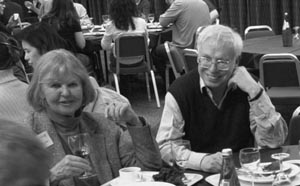
Beverly Griffin and Tomas Lindahl, 2002.
(Photograph courtesy of CRUK London Research Institute Archives.)
One of the first fruits of this decision was a paper describing the enzyme responsible for the adaptive response to alkylation first observed by Samson and Cairns. Tomas’s Mill Hill group showed in 1982 that the enzyme, O6-methylguanine-DNA methyltransferase, restored mutant O6-methylguanine bases to health by removing the methyl groups and disposing of them by methylating itself, the enzymatic equivalent of falling on a live grenade. Suicide inactivation, as the mechanism was christened, was not the only trick this enzyme had up its sleeve; subsequent work from Tomas’s lab showed that methylation at a different position in the protein turned it into a transcription factor that could not only activate its own gene, but also several others in the adaptive response pathway. Tomas is very proud of this work, published in 1986 in Cell: “If I may say so, I think it’s a well written paper and it had some impact! If I was going to mention one paper which I wrote myself … which I actually feel satisfied with, where I actually got things right, and I didn’t say too much or too little, it’s this one.”
Tomas clearly being a good researcher, Walter was very anxious to keep his new recruit happy and to involve him in the ongoing problem of what was going to happen to the scientists at Mill Hill. In 1982, Walter cut a deal that allowed the Mill Hill developmental biologists to move into a new, ICRF-funded unit at Oxford University, but the problem remained of what to do with Tomas. Shoehorning him into Lincoln’s Inn Fields was not an option that Walter, always an enthusiastic expansionist, favoured, and the healthy economic climate meant that there was money to spare for something more ambitious. The animal and service facilities at Mill Hill and Lincoln’s Inn Fields were going to be unified and expanded on the site of Clare Hall, an empty and decaying ex-smallpox and TB hospital near the newly built M25 motorway, so perhaps it would be possible to persuade Tomas to go there too?

Unlike John Cairns, who had thought that Clare Hall was far too isolated to work as a research laboratory, Tomas could see that there were possibilities out in the country, as long as several conditions were met. The first was that the laboratories should be big enough to accommodate a critical mass of scientists, which meant expanding the existing building plans, something Walter was happy to do. Owing to planning restrictions imposed by the local council, the laboratory could only be two storeys high at most, but this suited Tomas, who had observed in Sweden that “for some crazy reason scientists communicate very well horizontally and very poorly vertically. It sounds silly but for some reason a set of stairs is a really effective barrier, and walking down the corridor is much easier. And you can see that at Lincoln’s Inn Fields—the fifth floor and the fourth floor and the sixth floor were just different empires and didn’t actually chat. People on the same floor interacted closely and aggressively but at least interacted!” Accordingly, the Clare Hall laboratories took shape as an elegant linked collection of five low buildings, three devoted to research, and two to services, surrounded by landscaped gardens, all fitting rather well into the rural setting.
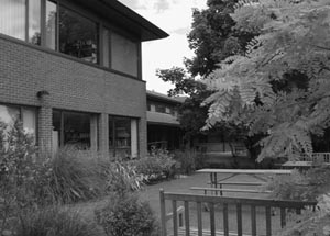
Clare Hall laboratories.
(Photograph courtesy of CRUK London Research Institute Archives.)
Having achieved the building he wanted, Tomas, now officially appointed Director of the Clare Hall Laboratories, decided that the labs would be very focused: “I was very concerned it wouldn’t be like the classical biochemistry or cell biology department that you see in universities, where because of the teaching, you have to be very broad like an old fashioned zoo—one zebra, one lion, one giraffe and one turtle and so on! That’s obviously an exaggeration, but if you have as your next door neighbour an expert on lipid metabolism it’s not perhaps somebody you easily share a seminar series with if you do nucleic acid or molecular biology work.” Not surprisingly, the focus that Tomas chose was his own—the biochemistry and metabolism of DNA, and the three Rs of Repair, Recombination, and Replication.
Sustained institutional success in science is a rare and interesting beast, seemingly dependent on a single factor: appointing the right person to be in charge. This certainly seems to have been the case with Clare Hall. Matched only by Cold Spring Harbor in terms of the scientific quality and impact of its publications, with its occupants clanking around under the weight of their many awards and honours, Clare Hall’s success revolved around its Director’s abilities. Tomas had an extremely good eye for hiring stars in the making, but turning Clare Hall into a place where the stars wanted to stay long term was an even greater feat.
As Director, Tomas was a figurehead that the Clare Hall inmates could trust and be proud of. His own work was good enough that he commanded respect from the faculty, and in return, he expected very high standards of scientific integrity and rigour; you were unlikely to do shoddy or boring work at Clare Hall and get away with it for long. Perhaps even more importantly, Tomas was genuinely interested in what everyone was doing and possessed an unusually high degree of intellectual generosity; he always had time to talk with other people about their work, even if there was no benefit to his own. This fostered a tremendous atmosphere of communal thinking, where ideas could be freely debated. A scientific problem shared is not necessarily halved, but the combined weight of a group of highly knowledgeable smart people thinking about something is guaranteed to eliminate half-baked ideas and unthinking dogma, clearing the way for original and exciting experiments. And people could afford to be generous with their thoughts; perhaps by luck as well as judgement, Tomas managed to hire people who were scientifically close enough to interact fruitfully but far enough apart that they were never direct competitors, which would have irretrievably poisoned the atmosphere.
Tim Hunt, who arrived at Clare Hall in 1990, eleven years before he won the Nobel Prize in Physiology or Medicine together with Paul Nurse and Lee Hartwell (who are to be found in Chapter 6), is an enthusiastic fan: “Tomas was so refreshing compared with the Professors of Biochemistry I’d known; here was a real biochemist who actually cared and knew what everything was. His whole philosophy was to try to reduce the energy barrier for doing experiments. He wasn’t at all manipulative or scheming. He was terribly pleased if you got a good result, although he was very hard on the people who didn’t! People didn’t last at Clare Hall if they couldn’t cut it.”
Tomas’s interest in his group leaders’ work extended to making sure that they were properly recognised for their achievements; thus, he was very active in nominating them for prizes and awards. He and the senior staff also made sure that junior lab heads with interesting work broke into the conference and seminar circuits as early as possible. This latter strategy was so successful that it gave rise to the mistaken impression that Clare Hall was a big institute; those not in the know assumed that the Clare Hall stars must be resting on a much larger bed of mediocrity, not realising that the stars were all there were.
A further boost to productivity was the almost complete absence of committees. Steve West, one of Tomas’s earliest recruits, characterises his managerial style thus:
Tomas was remarkable in that he never wrote an email. He would show up at the lab door and bring up something, then he’d listen to what you had to say—he’d never comment on it!—and then he’d go away and make a decision. You didn’t know whether or not you influenced that decision, because sometimes it wouldn’t be what you’d suggested, but he always made everybody feel that their opinion was valued, even if he completely ignored it! And that’s quite a good skill. And Tomas’s batting average was phenomenal in terms of making the right decisions.
Intellectual powerhouses cannot thrive long term unless their occupants are reasonably happy with their lot, and although Tomas’s egalitarian management style was greatly appreciated, he had two other invaluable assets, in the persons of Brenda Marriott and Frank Fitzjohn. With Tomas, Brenda and Frank formed a triumvirate that dealt with anything getting in the way of doing good science, and their importance to Clare Hall’s success cannot be overstated.
Frank’s job of Clare Hall lab manager was dreamt up by Tomas after his time on the Fifth Floor at Lincoln’s Inn Fields. In the frenetic atmosphere there, communal equipment was frequently broken, radioactive, or both, because nobody was prepared to take responsibility for fixing it. Anyone who has experienced the homicidal rage generated by standing in front of a broken centrifuge, holding a rack full of laboriously prepared and rapidly disintegrating samples, can understand the importance of having someone to ensure the smooth running of a lab, and Frank, in the nicest way possible, was a combined troubleshooter and vigilante without parallel. Bill House, who had hired him into the position, had simply described his job as: “Look after Lindahl,” and Frank took him exactly at his word. Whatever was required, from guiding fundraisers round the labs (elderly lady: “How many people work here?” Frank: “About half of them.”), to climbing into the drains to check them for radioactivity, fell within Frank’s remit. The move from Mill Hill to Clare Hall was seamlessly arranged and executed by Frank (Tomas: “Move us to Clare Hall, Frank, I’ll see you there in three weeks”), and thereafter, successive generations of scientists benefited from Frank’s efficiency (David Lane: “The good thing about Frank was, you asked him a question and he always said yes!”).
If Frank kept the place in perfect working order, Brenda Marriott, in the next office, was the guardian of its emotional well-being. Brenda’s potential had been discovered early on by John Cairns, who had hired her as a junior secretary when he first came to Mill Hill. After being roundly told off by the young Brenda for writing a “beastly” letter to someone, John realised that he had acquired someone rather out of the ordinary, and promoted her. By the time he left, Brenda was running the day-to-day business of the unit, and Tomas took her to Clare Hall as his chief administrator. Although she was as efficient in this as Frank was at managing the labs, she is fondly described by Tim Hunt as being the “den-mother” of the place, the person who turned Clare Hall into a family. Brenda was the person you first met as you arrived from the airport, the one who distracted your crying children as you struggled with your bags up the stairs to the flats in Clare Hall Manor, the grand 18th-century country house next door to the labs where incoming staff were accommodated. Brenda soothed broken hearts, found dentists for broken teeth, reined back the foolhardy and encouraged the nervous, settled disputes and gave advice, and generally acted as a conduit between the lab and Tomas.
Tomas describes his two lieutenants thus:
If Clare Hall became a success I can take some of the credit for that, but some of the credit is due to Brenda and Frank. They created [a] very efficient buffer, so when people wanted something, including lab heads, they would first go to Brenda and Frank, [who] had a very good sense when a scientist tried to take selfish advantage or when there was a real request.
Brenda is a very outgoing person, and people came and told her all kinds of things. She may have told me something like 5% or 10%, and she sifted the information I needed to know, because if she had told me everything, people would quickly find out and nobody would talk with her any more! There were things that perhaps I should have known that I didn’t know about, but more importantly, if there was something, she would come and say, do you know this, do you know that? And Frank was the same, and he also kept a good eye on the place, so if you found a dirty ultracentrifuge he would backtrack and find who had done that. Both he and Brenda had the sort of personalities that meant nobody on the site was keen to pick a fight with them or be told off by them. So they ran the place, and I was more the last instance when there was real disagreement.
Clare Hall wasn’t perfect. Undoubtedly, if people couldn’t or didn’t want to fit in, they were very unhappy there and left, along with those who didn’t make the grade. It wasn’t ideal for PhD students, because they really did feel the isolation and missed out on the metropolitan life being enjoyed by their compatriots at Lincoln’s Inn Fields. It spectacularly failed to appoint female group leaders, although Birgit Lane, one of only two women to run a lab there, charitably attributes this to benign neglect, rather than any more sinister motive. On the whole, however, it remains a shining example of how to create and maintain an environment in which intelligent, focused, important research can be performed as easily as possible.
Despite the stresses and strains of being Clare Hall Director, Tomas managed to go on working very productively. Uncovering the multitude of weird and wonderful enzymes involved in base excision repair was to take up much of his subsequent career, although he also made some notable diversions. One of these came to fruition in 1988, the year Tomas was elected to the Royal Society, when he and his postdoc Rick Wood made the first breakthrough towards identifying the UV-repair enzymes defective in xeroderma pigmentosum. Tomas promoted Rick into an independent position, and in 1995, Rick’s lab purified and reconstituted in vitro the human enzymes responsible for nucleotide excision repair. This tremendous feat gave Clare Hall the double in the excision repair biochemistry championships, because in 1994 and 1996, Tomas’s lab completed the equally laborious task of reconstituting base excision repair in vitro, using, respectively, the E. coli and human enzymes, some of which were remarkably conserved across the yawning evolutionary gulf.
In 2010, in recognition of his seminal contributions to the understanding of the biochemistry of DNA repair, Tomas was awarded the Royal Society’s Copley Medal, the oldest scientific prize in the world, whose other recipients include Michael Faraday, Charles Darwin, and, latterly, Stephen Hawking. However, by this time, the other inhabitants of Clare Hall were rapidly catching him up in the scientific eminence stakes. Two in particular, Steve West and John Diffley, have used the enlightened environment of Clare Hall to undertake ambitious, long-term projects, the anathema of the normal three-to-five-year grant cycle. Their stories, which complete this chapter, illustrate everything good about Tomas and the lab he built with such success.
Steve West and Genetic Recombination
The Clare Hall Laboratories of the Imperial Cancer Research Fund opened at the end of 1985, with a small but star-studded cast of five lab heads. Tomas was joined by Jeff Williams, the only one of the Mill Hill developmental biologists who had not wanted to move to Oxford, and new recruits David and Birgit Lane, who had been working at Imperial College and Lincoln’s Inn Fields, respectively. Jeff, David, and Birgit were well connected and greatly respected in their fields, and combined with Tomas’s reputation, their presence contributed greatly to the sense that Clare Hall was going to be something good. Furthermore, even though they were not really within Tomas’s original remit of DNA metabolism, they brought in some molecular biology skills that they were more than willing to teach to the biochemists.
Steve West, the last group leader to make up the new Clare Hall intake, had not intended to like Clare Hall at all, having only come for an interview from his then workplace, Yale, because he fancied a free trip to visit his mum in Yorkshire. However, almost three decades on, he is still an inmate, one of the big beasts of the DNA repair world, and probably Tomas’s most inspired hiring. As well as all the normal attributes that a great scientist requires, Steve’s reputation rests on his eye for major problems, his extraordinary skill as a biochemist, his phenomenal workrate, and his tenacity. All of these are exemplified in a knotty problem that Steve brought back with him from the United States, and that in the end took 25 years to solve: how does the cell cut through the tangled products of genetic recombination to regenerate two functional double helices?
Until the 1960s, genetic recombination was viewed by most biologists as an esoteric problem worked on by a small number of fungal geneticists. The exchange of information between two strands of DNA is essential for evolution and happens every time two people get together to reproduce, but pre–Watson and Crick, its study was caught in an intimidating briar patch of statistical analysis and complicated hypotheses. However, like everything else in biology, the DNA revolution meant that the geneticists’ ideas could be reformulated in terms of the interaction of DNA molecules, opening the way for biochemists and molecular biologists to start thinking about exactly how recombination worked.
Recombination involves the breaking of a DNA double helix, the invasion of one of the strands of the broken helix into a homologous (identical) sequence, generally on the other partner chromosome, a period in which both chromosomes are joined together in a complicated four-way knot called a Holliday junction, and the cutting of the knot to resolve the tangle into two discrete double helices. Before resolution, the Holliday junction can move along the DNA strands, creating a situation in which, as if in an elaborate barn dance, strands can switch partners, ending up, when the molecular music stops, on the other chromosome from which they started. This process of branch migration, during which genes or parts of genes cross over from one parent’s chromosomes into the other’s, creates the concoction of mixed-up inheritance known on a small and delightful scale as a new baby, and on a larger scale as the basis of genetic diversity.
Early on in the history of recombination research, it became evident, from work performed by Dick Boyce and Paul Howard-Flanders, the codiscoverers of excision repair, that homologous recombination was also very important for repairing DNA damage. The most dangerous hit a DNA molecule can take is one that breaks both its strands simultaneously, because there is then no template from which to repair the lesion. Like a snapped rope, the flailing strands can be spliced to anything else that happens to be around, or simply left dangling, and the consequences to the cell can be disastrous: Large- and small-scale mutation and problems with replication lead inexorably to the catastrophes of cell death or (in multicellular organisms) cancer. Homologous recombination is one of two mechanisms for fixing double-strand breaks and averting these unpleasant fates.
In 1965, Alvin Clark and Ann Dee Margulies, working in Berkeley, published a set of E. coli mutants defective for both recombination and repair of UV damage, one of which they named RecA. By the mid-1970s, it had become clear that whatever the RecA protein did, it was central to both recombination and repair. A great deal of effort was expended to discover its identity, culminating in 1977 with the publication of three papers detailing the purification of RecA protein. One of this trio was an early appearance in print of Steve West, then a young lad from Hessle, near Hull, doing a PhD whilst supplementing his income DJ-ing round the student haunts of Newcastle.
Steve West’s dad was a fish buyer in Hull, and despite parental expectations, Steve knew very early on that he did not wish to follow his father into a job at the fish docks: “He had a big red face, typical of someone who works outside in the cold, and his hands were … well, you can imagine! I can remember when he came home he stank of fish, and had fish scales all over him, and I thought, ‘Nooo, that’s not for me.’ ” Instead, Steve became the first in his family to go on to higher education, spending three years at Newcastle University to earn an undistinguished degree in Biochemistry: “I had too many other interests, and I could never be bothered to learn all the stuff, so I was never very good at exams. I was absolutely crap at maths too.” Despite his unpromising record on paper, Steve got a PhD place at Newcastle with Peter Emmerson, an ex-postdoc of Paul Howard-Flanders, and realised that finally, he had found something that fired his enthusiasm: “I got in the lab and life was easy! You don’t have to learn anything, and I always knew what the next thing to do was. I was very good at the bench and I was willing to put myself out to do experiments that would take a long time. I was obsessive about it, discovered what I liked doing.”
Steve’s project was to work on a mystery protein, unoriginally called Protein X, which appeared in vast quantities following irradiation of E. coli. It was not a subject of great interest to his supervisor: “Peter was famous for saying: ‘Protein X is for the birds—why are you bothering with this stupid thing?’ ” but Steve hung on, reckoning that if a cell made something in such large amounts, it had to be important. He was more than vindicated when he discovered that Protein X was in fact RecA, and the 1977 paper describing this finding set Steve well on his way in the world of DNA metabolism. He moved to Yale to the lab of Paul Howard-Flanders, who turned out to be an understated Englishman with an almost pathological disinclination to dictate his new postdoc’s research direction. Under the circumstances, the easiest path for Steve was to simply continue with his RecA work, which in any case was still very interesting, so this he duly did.
Steve West, 1978.
(Photograph courtesy of Steve West.)
Life at Yale was very happy for a single-minded biochemist: “I stayed all night, worked fifteen hours a day, seven days a week. I used to take my girlfriend to a Greek restaurant near the lab, and the nice thing was, I could nip out between courses and change dialysis buffers, and she could stay there and go for a dance—it was one of those little restaurants where they do Greek dancing—until I came back from changing the buffer. She thought the mad scientist thing was cute.” Much to Steve’s good fortune, this amazingly tolerant person eventually agreed to marry him.
Steve published 20 papers during his seven years at Yale, in the process converting his boss, who had started life as a radiobiologist, into a confirmed believer in biochemistry. The RecA field was incredibly competitive, with several labs, including another one down the corridor at Yale, trying to figure out what RecA did during recombination, but at the tail end of 1980, Steve hit the jackpot. He was looking at what happened when purified RecA was mixed with two separate DNA molecules, which he was able to distinguish because he had tagged one with radioactive tritium and the other with 32P. Instead of seeing changes in the sizes of the DNA bands, or perhaps the appearance of a new one, Steve got a most peculiar result; the tritium and 32P labels seemed to have got mixed up between the two DNAs, so that both fragments contained both labels. After a couple of repeat attempts, during which the mixed up labeling stubbornly persisted, Steve set the whole experiment up again on Christmas Eve. He relates what happened next:
I came in on Christmas morning, and half the tritium was still in the 32P, and half the 32P was still in the tritium. So I called Paul at home at lunchtime on Christmas Day, and I said: “I’ve got this really weird result and I just don’t understand it.” … And there was this absolute silence on the phone, like a minute. Paul was actually a little strange in that he would have these speech gaps, he would stop talking for some time, but this was abnormally long. So I’m sitting there, and a minute goes by, and he said: “Well Steve, if it’s a recombination protein, maybe that’s what it’s supposed to do.” And at that point I realised it had done strand exchange! And then we were both kind of silent to each other on the phone, because it had done strand exchange—it had done something which was miraculous. And that was cool—a cool moment.
RecA was, indeed, the catalyst for strand exchange, coating the loose end of a DNA molecule to create a nucleoprotein filament, searching around for a homologous partner, pushing its double helix apart, and feeding the broken DNA in to start the process of recombination. For the next three years, Steve pretty much published whatever experiment he did, as the field leapt forward, armed with the knowledge of RecA’s function. By 1983, Steve had worked out conditions in which he could take his purified RecA, add it to two different plasmids in vitro, and initiate strand exchange. Then, by adding some DNA polymerase and DNA ligase (the enzyme that glues loose ends of DNA together), he could magically transform the two circular plasmids into a single, figure-of-eight molecule joined at its waist by a Holliday junction. If put back into an E. coli strain that lacked the RecA gene, such figure-of-eights were resolved into a mixture of products that could only have arisen by homologous recombination. This elegant experiment, published in Cell, unequivocally showed that Holliday junctions were really the recombination intermediates, which had been suspected but not definitively proved, and also showed that RecA was not required for the final junction resolution step in the recombination reaction.
At this point, having been at Yale for five years, Steve knew it was time to go on the job market, for which he needed an interesting project that was entirely his own. The mechanism of Holliday junction resolution was a complete black box, so, being nothing if not ambitious, Steve decided that he would go hunting for the enzymes involved, with the goal of getting the whole of homologous recombination to work in vitro. In his last two years in Paul’s lab, he set up the basics for the resolvase project, doing the lab three-week preps to make figure-of-eights, cracking open bacteria to make protein extracts, and seeing if any of the extracts could resolve the figure-of-eights in vitro. Much to his credit, Paul funded all of the work, but wanted none of the glory, a practice that Steve maintains: “He was fabulously generous and I try to do the same with people in the last year they’re with me—do what you want, I don’t care, and I don’t want to be on the paper. And it’s good, it allows them to have their own independence.”
Despite having some good job offers from places in the States, Steve was seduced into coming to Clare Hall after meeting Tomas at an end-of-conference banquet in Cambridge:
It was a nice evening and Tomas likes his wine, so I think I drunk far, far too much! A couple of weeks after I got back he sent me a letter which said: “We’re building this place at Clare Hall, and would you like to come and have a look at it, and see what we have to offer?” I actually had no interest at all, because I’d been in the States for seven years, my girlfriend, who became my wife, was American, and we had no intention of coming back here. But my mother still lived here, so I thought, free trip back, go and see Mum. So I visited my Mum, and then came to Clare Hall, which was still a hard hat site. Tomas was pretty astonishing. He said: “If you come here we’ll give you the resources—a technician, a graduate student, a couple of postdocs after a year or so, you don’t have to write grants, and you can decide where you want the benches to go, where you want the sinks to be.” He actually said those golden words: “If you can’t succeed here, you probably won’t be able to succeed anywhere,” which was very clever actually, because when you’re starting a lab you’re scared to death you’ll fail.
Steve duly arrived in his empty lab space in 1985 and got on with his resolvase-hunting experiments. Things had become a little easier since he’d begun them, because the three-week figure-of-eight preps had vanished into the past, in favour of sticking together four radioactively labeled artificial oligonucleotides (short lengths of DNA, in this case, 60 nucleotides long) to create a cruciform structure that was a perfect in vitro mimic of a Holliday junction. With this innovation, introduced by Nev Kallenbach at the University of Pennsylvania in Philadelphia, experiment times were reduced to afternoons, rather than weeks, and you could tell whether the junction had been cut simply by running the reaction products on a gel and seeing whether any of the original oligonucleotides had changed size from 60 bases long to 30. By 1989, Steve and his first postdoc, Bernadette Connolly, could definitely get resolution of the cruciforms by adding E. coli extracts to them, but try as they might, they simply could not purify the responsible enzyme from the bacterial protein soup. At this point, having exhausted the heavy guns of biochemistry, Steve decided to deploy the more subtle weapon of genetics, calling up his mate Bob Lloyd, who was one of the best geneticists in the field. Steve and Bernadette had already dabbled with making protein from known bacterial recombination mutants, to see if any of them were defective in resolvase activity, but all had been able to resolve cruciforms with no problem, showing that they must still contain functional resolvase. However, Bob Lloyd had four new mutant E. coli strains—ruvA, ruvB, ruvC, and orf26—which he was happy to send down from his lab in Nottingham to Clare Hall.
In the last experiment he did himself before retiring from the bench, Steve made cruciforms, added extracts from each of Bob’s mutant strains, and ran the diagnostic gels. Using normal, ruvA, ruvB, and orf26 extracts, he got the expected 30-nucleotide-long resolution product, but with the ruvC extract, there was a complete blank at the expected place on the gel. It looked as though ruvC might be the elusive resolvase, and this was rapidly confirmed by getting a ruvC-overexpressing strain from Bob Lloyd and showing that it was superefficient at cruciform resolution. In 1991, the West and Lloyd labs published a Nature paper describing the purification of the RuvC resolvase, which they showed was a nuclease able to sit on Holliday junctions and cut two opposing DNA strands symmetrically.
Steve’s lab was now on a roll and showed very quickly that the RuvA and RuvB proteins were responsible for branch migration, acting together as a molecular motor that could move Holliday junctions along DNA until the RuvC protein, squatting on top of them, found a sequence that it liked, and cut the DNA to resolve the junction. Thanks to his work, and that of a few other labs, notably that of Hideo Shinagawa in Osaka, the mechanics of homologous recombination in bacteria were pretty much sorted out, and not a moment too soon; research into DNA repair and homologous recombination was about to become extremely trendy.
The sudden upturn in interest came about after two key findings. Firstly, the machinery of DNA repair and recombination was shown to be evolutionarily conserved to a remarkable degree, such that close relatives of many of the bacterial enzymes were found in mammals, and, secondly, mutations in the repair machinery started to crop up in cancers, beginning with the discovery in 1993 that a form of hereditary colon cancer was caused by defects in the repair of mismatched nucleotides. The tentative connection between DNA repair and human health, first made by Jim Cleaver in his xeroderma work, had finally clicked firmly and irrevocably into place.
Steve, somewhat unwillingly, was dragged out of bacteria into mammalian cells, although the Clare Hall facilities made the transition less alarming:
It was postdoc pressure! Postdocs would join the lab, and I don’t think they saw their future in bacterial proteins—they wanted to go into mammalian systems and find the same things. So the lab evolved from bacteria into human cells, probably over two years. The beauty was having Cell Services here—rather than growing E. coli, they would grow us mammalian cells, and we were just making extracts, so what’s the difference? Extracts are extracts and you do the same experiments. So the transition was invisible—there was no real decision to be made, and the lab evolved from working on bacteria to human systems. Mind you, it would never have happened without Cell Services, because I’ve never grown a cell, and if I had a lab in a university, and had to grow cells in my own lab, I might have never made that transition.
As well as making a start on the search for the mammalian version of the RuvC resolvase, Steve had not forgotten his first love, RecA. In 1994, his lab was the first to purify its human homologue, RAD51. RAD51 coats single-stranded DNA to create a nucleoprotein filament that invades a homologous double helix, starting the process of recombination in a very similar way to RecA. The subsequent discovery that RAD51 was both positively and negatively regulated by the BRCA2 protein in response to DNA damage brought this work to centre stage in cancer biology. BRCA2, like p53, is a tumour suppressor, essential for guarding the cell from possibly oncogenic mutations. Without it, homologous recombination is defective, and double-strand breaks can only be repaired by a more error-prone mechanism called nonhomologous end-joining, leaving the cell extremely vulnerable; that mutation of BRCA2 is found in some 10% of inherited breast cancers is therefore no surprise.
As the West lab grew and diversified, they continued to take on and meet multiple biochemical challenges in mammalian cells, all relating to defects in DNA repair and their effects on disease. They worked out that the huge BRCA2 protein, a nightmare to handle because it is so big and likes to fall apart, controls the ability of RAD51 to bind DNA, and they sit at the forefront of research into repair-linked diseases such as Bloom’s syndrome, Fanconi Anaemia, and the unpleasant progressive neurological condition Ataxia with Oculomotor Apraxia 1. For all these achievements, Steve has won many awards and has been a fellow of the Royal Society since 1995. However, through all the years of success, Steve carried another project close to his heart, a failure-ridden enterprise that, despite its promising start, was foundering in a sea of dashed hopes and postdoctoral frustration: the quest for the mammalian resolvase.
To begin with, things had gone very well. As early as 1990, Steve’s first graduate student, Kieran Elborough, was able to detect resolvase activity in extracts from mammalian cells, but as the decade wore on and the Cell, Nature, and Science papers rolled in for everything else, the resolvase project stuck fast. The problem was in the purification: “If you purified it, you’d lose activity, and then the strangest thing in the world would happen—the activity would come back. And we know now it’s all to do with post-translational modification—it’s inactive when it’s phosphorylated; it’s activated by dephosphorylation. So it would be coming off a column with other things that would phosphorylate and deactivate it. You’d take that, fractionate it again and lose the phosphates, and the activity would come back again. It was so frustrating. SO frustrating!”
The century turned over, and Steve was elected to the Academy of Medical Sciences and won the Leeuwenhoek Prize of the Royal Society, capping that in 2008 with Europe’s most prestigious scientific award, the Louis-Jeantet Prize for Medicine. The resolvase project still languished in the doldrums and had even lost its sad little entry in the London Research Institute Annual Report. The project was so unsuccessful and such hard work for people that it gained an unhealthy reputation as a postdoctoral graveyard. In the end, Steve had to let it go fallow for two or three years because he simply couldn’t talk anybody into trying it.
By 2005, Steve was getting pretty desperate: “It was funny, people said to me: ‘What if after all these years somebody else finds it?’ and I said: ‘Fantastic! I just want to know what it is now, I don’t care!’ ” However, salvation was at hand, in the shape of Stephen Ip, a new postdoc from Hong Kong who was even fonder of hard work than Steve himself and was, in addition, willing to take on a risky project if his supervisor insisted. Together with a second postdoc, Uli Rass, Steve and Ip formulated a last-ditch, two-pronged assault on the resolvase problem. Uli, subsequently joined by Miguel Blanco, would perform a massive screen using a molecular library containing every gene from the yeast Saccharomyces cerevisiae. The genes all came attached to a TAP-tag, a clever modification by which the proteins encoded by the genes could be easily hooked out of cell extracts. The plan was to individually purify all of the yeast proteins with unknown functions and test them one by one for resolvase activity using cruciform substrates. There were about 2000 to work through. While this was going on, Ip would make extracts from 200 L of the human cancer cell line HeLa; fractionate the extracts by protein size, charge, and chemical behaviour; and test the individual fractions for resolvase activity in the cruciform assay. Once the fraction with the most activity had been identified, the proteins in the fraction would be further separated according to size by running on gels. The size-separated samples would be tested again for resolvase activity, and, finally, the proteins in the sample with maximum resolvase activity would be identified by their mass-to-charge signature on a mass spectrometer. The whole process was as expensive as it was laborious, and no sane grants committee would have sanctioned it, so it was just as well that the Clare Hall core funding from Cancer Research UK was picking up the tab.
Uli slowly worked his way through the TAP-tagged yeast library, whilst Ip got on with the brute-force purification. And, finally, after 1100 TAP-tagged protein assays and Ip spending so long in the cold room running samples that he could have walked to the Antarctic and back, there came an afternoon in 2008 when Steve looked up from his computer and saw his heroic resolvase postdocs framed in the doorway of his office: “Uli, Ip and Miguel stood at my door and there were tears running down Ip’s cheeks, because they knew they had it!” Almost simultaneously, Uli had isolated the yeast resolvase, Yen1, and Ip had found the human one, GEN1. Amazingly, they were homologous to each other but were entirely unrelated to RuvC, their bacterial counterpart. The 20-year journey from E. coli to humans had ended at last.
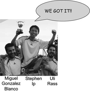
The three resolvase postdocs.
(Photograph courtesy of Steve West.)
There would be many more twists and turns to come in the resolvase story, but for Steve, finding Yen1 and GEN1 was one of the biggest thrills of his career, easily up there with his Christmas morning realisation that RecA could do strand exchange: “It was a fantastic little story. It’s fantastic just in terms of the ICRF and Cancer Research UK, because you could not have done this anywhere else in the world. You could not find any other funders who would fund the same project for twenty years. You had to be able to do this sort of thing without justifying it—it was at the point where it was becoming unjustifiable just to keep doing it.”
And the future? “Now we know there are three to four pathways of resolution inside eukaryotic cells, of which GEN1 is one. It’s fascinating because it turns out that the resolvases are controlled by post-translational modifications. So each is active at a different time in the cell cycle. Understanding what these proteins do, and how they are regulated and interact, should keep me going till I pack it in. I hope so, anyway!”
John Diffley and DNA Replication
In 1990, David and Birgit Lane were lured away from Clare Hall by the University of Dundee, giving Tomas an opportunity to hire new faculty, but also a problem. The loss of the Lanes triggered an outbreak of mingled pessimism and Schadenfreude in some parts of the scientific community; how would such a small, quiet place recover from the departure of two of its biggest guns? The notion that Clare Hall was doomed was soon scotched, however, by the surprise announcement that one of biochemistry’s most visible and popular figures was moving there. Tim Hunt, who, most people thought, would only leave his beloved Cambridge when carried out feet first in a wooden box, had decided to abandon the creaky environs of the university Biochemistry Department for somewhere more congenial and better equipped, where he could concentrate on research rather than teaching. Tomas was delighted with his coup: “I was obviously very happy to take on somebody with Tim’s high reputation. It gave me some personal satisfaction when people said to me, ‘Oh dear, who are you going to find to replace David Lane?’ and I was able to say ‘Tim Hunt!’ That really shut them up—none of them expected that answer!” Tim was equally pleased with his decision: “It was just wonderful suddenly to be surrounded by all these biochemists. Steve West was a total revelation to me when I went there. I was amazed—here was this guy putting recombination on the biochemical map. Sydney [Brenner] was always interested in recombination, but it was always phage genetics and that sort of thing—very very hard to understand.”
After Tim’s arrival, his contagious enthusiasm and boundless capacity for discussing even the most eclectic scientific subjects became one of the most energising aspects of Clare Hall life. When, much to his shock and amazement, he was awarded his 2001 Nobel Prize for the discovery of cyclins, the key regulators of the cell cycle, Tim’s presence added a touch of star quality as well. His office became a high point of Frank Fitzjohns’s Clare Hall supporters’ tours, as Brenda Marriott recalls: “Tim was wonderful with the appeals department. Frank would take the tours round that way and Tim would come to the door and stand there and smile, and after Frank told the tours who he was, he’d say very grandly, ‘You can touch me if you wish!’ And they would all start giggling!”
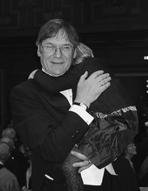
Tim Hunt and daughter, Nobel Prize ceremony, 2001, Stockholm. (Photograph courtesy of Tim Hunt.)
To complement Tim’s interest in the cell cycle and to steer Clare Hall closer to his original ideal of a pride of DNA-focused lions, Tomas’s other recruit, John Diffley, came from Bruce Stillman’s lab at Cold Spring Harbor, which remains one of the major centres for research into the biochemistry of DNA replication. John is from Brooklyn, although he spent his school years in Suffern, New Jersey, where he attended a single-sex Catholic high school. At 16, his life changed radically when he got into New York University (NYU) as a premed. According to John, moving from New Jersey to university digs in Greenwich Village was “great, for all the wrong reasons and all the right reasons,” but it didn’t take long for him to realise that although New York was a great place to be, he had no desire to be a medic there or anywhere else: “During the first summer I worked in a hospital and realised I never ever wanted to do medicine—I hated it!” Instead, John enrolled at the age of 20 in the PhD programme at NYU to work with a junior group leader, Chris Brakel, who was interested in DNA replication. After a year, however, disaster struck: Brakel decided that she was going to leave science, and John was left high and dry, in possession of the last year of Brakel’s grant money, but without a PhD supervisor. Undaunted, he wangled himself into another lab at NYU belonging to Mike Kambysellis, a Greek Drosophila geneticist, splurged the remnants of the Brakel grant on enough biochemistry supplies to keep himself afloat for the duration, and got on with his original project, purifying the main Drosophila replicative DNA polymerase. It was a crash course in independent research; nobody else was doing any biochemistry in the Kambysellis lab, and to cap it all, the lingua franca was Greek—“they’d have these massive Greek arguments—quite amusing!” So John was pretty isolated: “I never went to a meeting or anything. There was nobody to tell me to do this or that or to give me any guidance. I had to do it on my own, figure out what I wanted to do. But I loved it; I completely loved it. I can’t think of a better thing to do. I guess biochemistry is really puzzle solving—what most people love about science—figuring out how something works when you don’t know.”
Having had to figure out his next career move by himself as well, John got into Bruce Stillman’s lab on the strength of the two extremely competent papers on polymerase purification that he published during his PhD. Any normal person, especially one coming out of John’s situation, would probably have taken advantage of the new lab’s expertise and done a solid biochemical project on DNA replication. However, John is nothing if not unusual: “I started doing yeast genetics, and again I taught myself. Bruce is a great biochemist but he doesn’t do yeast, so I set up yeast in the lab. Everyone else was doing SV40 replication and I was doing yeast—I guess there’s just a level of contrariness in my personality!”
Unsurprisingly, in the 30-odd years since Kornberg had found the first DNA polymerase, an awful lot had been discovered about the enzymes responsible for replicating DNA: Helicases unwound the DNA double-helix; primases started the new strands off; multiple types of polymerase, tailored to particular tasks, and each equipped with an editing function to cut out mismatches, got on with elongation; sliding clamps held the replicating strands of DNA steady; and ligases sewed the new pieces together to make the final product. The whole process resembled a busy factory production line, and although there was still considerable interest in the individual components of the process, attention was shifting to finding out how it was regulated. What was responsible for triggering replication, and how does the cell know to switch the replication machinery on once, and only once, during the cell cycle?
The Stillman lab was almost alone at the time in studying initiation, the first step in the replication process, in eukaryotic cells. As John Cairns had shown in the 1960s, bacteria, with their small genomes, start replicating from one particular point in their DNA, known as the origin of replication, and just continue until they have gone full circle. This is a relatively simple process. Eukaryotes, however, have a far larger complement of DNA, which is broken up into separate chromosomes, and therefore they need multiple origins of replication in order to perform the process on each chromosome, and within a reasonable timescale; in the case of mammalian genomes, origins of replication number in the tens of thousands. The problem that John wanted to tackle was how all these origins were able to fire just once in every cell cycle to orchestrate the complete replication of the genome, and, to begin with, he needed to define how the origins were recognised by the replicative machinery. During his time with Bruce, he purified two proteins that bound to yeast replication origins, and it was on the back of this, and other work in the lab’s adenovirus model systems, that Tomas hired him.
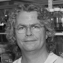
John Diffley, ca. 2010. (Photograph courtesy of CRUK London Research Institute Archives.)
Like Steve West before him, John applied to Clare Hall for less than scientific reasons; he fancied a trip to England, where he’d never been, and the ICRF job advert he’d seen in the back of Nature would pay his airfare. However, once he got here, the Clare Hall magic took over. Tomas remembers the stringent recruitment process very well: “I think I took him to the local pub and it was Sunday afternoon, and he was sitting looking out at the slopes of the ridge up there, a very nice British early summer day.” John met the other group leaders, liked them, liked their work, liked the extremely generous starting offer, and decided to take the job: “The similarity to Cold Spring Harbor in terms of isolation is what appealed to me when I came here. I saw a lot of the same things in it and it either suits you or it doesn’t. I did miss the people passing through Cold Spring Harbor at first—I started having to read the literature again! When you’re at Cold Spring Harbor you don’t have to read anything as you find out about it.”
Following John’s arrival, Tomas was slightly dismayed to find that his new recruit had, temporarily at least, abandoned biochemistry. Taking his example from the transcription field, where people were having a lot of success in reconstituting transcription in vitro by simply adding cell extracts to DNA templates, John had spent the end of his postdoc trying to do the same with replication but had completely failed. It was a perfectly sound strategy, but what he didn’t know at the time was that replication is so tightly controlled that most of the proteins involved are either absent or held in an inactive state until immediately before they’re required; adding random extracts was never going to work. In the face of this defeat, John decided to develop some approaches to look at what was sitting on the yeast origins of replication in vivo, in the cell itself, and for this, he had to use an extremely fiddly technique called genomic footprinting.
The principle of genomic footprinting is simple: if proteins are bound to DNA, the DNA is protected from attack by nucleases. Therefore, if you open cells up very gently, making sure not to disrupt the protein–DNA interactions, and then use nucleases to digest the DNA, the place where your protein of interest is bound shows up as a larger, undigested fragment of DNA. This can be visualised by the usual method of adding huge amounts of radioactive 32P to label up the DNA, and running it on a gel afterwards. In practice, the experiment is extremely difficult to perform cleanly; it’s really fiddly, with multiple steps, so there are many opportunities to lose material, which is a big problem because you are dealing with vanishingly small amounts of DNA in the first place. Furthermore, at the end of all your labours, you are often confronted with a complete mess, with so many background bands on the gel that the footprint is uninterpretable. It took John and his PhD student Julie Cocker almost three years to troubleshoot, during which time Tomas got progressively more worried: “It was clearly a very difficult project, and it was clear he wasn’t getting anywhere with it. So I had a talk with him to see if he was sure it was what he should be doing. He felt the solution was just around the corner and he was right. But I did pay attention that way if somebody was trying something that was too ambitious.”
The result of John’s departure from biochemistry was a Nature paper in 1992, identifying a complex, called the origin recognition complex, or ORC, that was bound to yeast origins of replication in real time in the cell, as well as in a test tube. However, there was a lot more to be done. Although ORC was clearly sitting on origins of replication, it was unlikely to be what determined when those origins fired during the cell cycle; it was bound to DNA almost all of the time, and the protein that flicked the “on” switch was likely only to be there right before it was needed.
John’s lab expanded their footprinting operations and found that one particular DNA sequence in yeast replication origins was only occupied by proteins during a phase of the cell cycle called G1, which occurs immediately before S phase, the point where the genomic DNA is replicated. The new data appeared in a major paper in Cell that showed that this protein complex, dubbed the Pre-Replicative Complex (preRC), was very similar to Xenopus Licensing Factor, an activity in frog eggs that was required for initiation of replication. That similar complexes were found in two such dissimilar organisms suggested that the control of replication regulation might be broadly the same in all eukaryotes, and this has, indeed, turned out to be the case.
With the genomic footprinting working well, John deployed his other nonbiochemical skill, yeast genetics, to identify mutants unable to assemble the preRC properly. Using these mutants, he could bootstrap his way to the proteins they encoded and start purifying them, with the aim of reconstituting the preRC in vitro, finally returning to his roots as a biochemist. John had already started a very successful collaboration on the ORC with the yeast molecular biologist Kim Nasmyth, and in 1996, the two labs published a joint Nature paper, identifying the first preRC complex protein, Cdc6.
The preRC is the thread running through all of John’s subsequent research, and 18 years on from the first in vivo footprint, John’s lab finally achieved its in vitro reconstitution. This scientific and technological tour de force put a vital piece into the puzzle of how replication is limited to a one-time event during the cell cycle, because regulation of the preRC turns out to be the most important thing. The cell has evolved an elegant system for preventing origins from firing more than once: only allow the preRC to be assembled at a particular time in the cell cycle, and only allow its subsequent activation during S phase if the cell is confident that all systems are go.
The first step, preRC binding, now called licensing, is regulated so that it can only occur in the early stages of the cell cycle, from just after the cell has split in two (mitosis) until the end of G1. The ORC, together with Cdc6 and another protein, Cdt1, uses the cellular energy source ATP to load a helicase motor, comprising another six proteins together known as Mcm2–7, onto origins. The preRC complexes sit on the origins and wait. Although they are ready to go, they need the biochemical equivalent of car keys to get their engines going, and these are only provided by the cell during S phase, in the shape of specific activating enzymes that are not made in any other stage of the cell cycle. The enzymes, which belong to a group called cyclin-dependent kinases (CDKs; see Chapter 6 for more about how they work), turn on the Mcm2–7 helicase, which starts to move along the DNA, unwinding it as it goes so that the rest of the replication machinery can load and begin synthesis. After the genome has been replicated, the cell divides, and the whole procedure starts again.
The processes of assembling the preRC on DNA and getting it activated are so crucial to correct replication that there is layer upon layer of regulation of both. Working out the intricacies of how the cell achieves its aim of perfect reproduction of the genome has taken John into the fields of transcription, whose enzymes have had to learn to share nicely because they use the same template as the replicative machinery, and, no doubt to Tomas’s satisfaction, into DNA repair territory. To start DNA replication, the cell has to pass a checkpoint, where DNA damage is detected and reported to the replication machinery. A certain amount of careless, error-prone replication is allowed in the interests of keeping the S-phase show on the road, but if things look really bleak, the cell calls a halt to the whole process and initiates a frantic repair programme before allowing S phase to continue. Any origins that have not yet fired are inactivated by switching off the Mcm2–7 helicase, and the replication machinery sitting at stalled replication forks is stabilised, until it receives the signal to resume synthesis.
Such complexity of regulation might appear daunting, but to a biochemist of John’s calibre and ambitions, the thought of purifying 60–70 proteins and then assembling them into somewhere around 25–30 complexes is his idea of the ultimate good time:
This may be a bit of a boy thing, but ever since I was a little kid I took things apart. I broke every toy I ever got for Christmas within a week because I took it apart and couldn’t put it back together. So now that I’m finally putting things back together, it’s great! There are really interesting questions to ask about how these things work. What’s particularly interesting is that there isn’t a precedent in viruses or in bacteria—it really works completely differently from replication in any other realm of life as far as we know, so the things we’re going to learn are really new. Replication is the centre of everything in the nucleus, because everything that happens is coupled somehow with replication, whether it’s chromosome inheritance or sister chromatid cohesion, so I think having an in vitro replication system is really going to open up a lot of stuff for biochemical analysis.
In the short term, the loading of the Mcm2–7 helicase onto the origin is currently occupying John’s thoughts because persuading a multisubunit helicase to wrap itself round DNA is an intriguing topological problem. In addition, his lab has just found that ORC, which together with its accessory protein Cdc6 is the ATP-burning enzyme that loads the helicase up, can tell whether the helicase is intact or not, and unload it if components are missing. The ORC ATPase, unsurprisingly, also has a further level of regulation, by CDKs.
In the long term, John’s objective is simple: “We’re close to having the whole replication process in vitro. We’ve got about a third of the proteins purified, but in yeast we know all the proteins involved and it really just means purifying all the proteins and reconstituting them. But my real goal is that I want to reconstitute a yeast mini-chromosome with replication and a gene, so we can look at interactions between replication and transcription. I think that’s a feasible thing.” His old boss, Bruce Stillman, agrees: “The extraordinary clarity of his observations, the crispness of his data, and the beautiful logic of his ideas and discoveries have been characteristics of John’s research. He has always addressed important problems and has been at the forefront of solving them. I would venture to say that John is one of the best scientists today who focusses on how our genome is inherited from one cell generation to the next” (Stillman 2003).
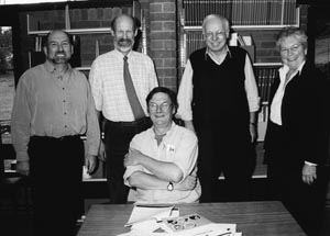
Nobel Prize celebrations, Clare Hall, 2001. (Left to right) Steve West, Frank Fitzjohn, Tim Hunt, Tomas Lindahl, Brenda Marriott. (Photograph courtesy of Steve West.)
Web Resources
www.nobelprize.org/nobel_prizes/medicine/laureates/2001/hunt-autobio.html# Unfortunately for this author, Tim Hunt’s Nobel Prize was won for work conducted in Cambridge and Wood’s Hole and therefore falls outside the remit of this book. To read more about him and his work on cyclins, a good starting point is the Nobel website.
www.youtube.com/watch?v=lZxc3Pohf70 Good video about DNA structure.
www.youtube.com/watch?v=cvU4kEMvbm4 Fantastic video of Matt Meselson describing “the most beautiful experiment.”
www.youtube.com/watch?v=SROcb5h1–w Useful animation of a Holliday junction.
Further Reading
Cairns J. 1997. Matters of life and death. Princeton University Press, Princeton, NJ.
An extremely readable account of the things John Cairns thinks are important about science, mingled with a bit of autobiography in passing.
Diffley JF. 2011. Quality control in the initiation of eukaryotic DNA replication. Philos Trans R Soc Lond B Biol Sci 366: 3545–3553.
Friedberg E. 1997. Correcting the Blueprint of Life: An historical account of the discovery of DNA repair mechanisms. Cold Spring Harbor Laboratory Press, Cold Spring Harbor, NY.
I am indebted to this simultaneously clear, entertaining, and erudite account of the early days of DNA repair—well worth searching for.
Kornberg A. 2000. Ten commandments: Lessons from the enzymology of DNA replication. J Bacteriol 182: 3613–3618.
Lindahl T. 1996. The Croonian Lecture 1996: Endogenous damage to DNA. Philos Trans R Soc Lond B Biol Sci 351: 1529–1538.
West SC. 1996. The RuvABC proteins and Holliday junction processing in Escherichia coli. J Bacteriol 178: 1237–1241.
Quotation Sources
Bruce Stillman remarks on John Diffley for Paul Marks Prize for Cancer Research 2003 Prize Winners, Memorial Sloan-Kettering Cancer Research. www.mskcc.org/research/paul-marks-prize-research/2003-prize-winners.
Cairns J. 1963. The bacterial chromosome and its manner of replication as seen by autoradiography. J Mol Biol 6: 208–213.
Cairns J, Stent GS, Watson JD. 2007. Phage and the origins of molecular biology: The centennial edition. Cold Spring Harbor Laboratory Press, Cold Spring Harbor, NY.
Friedberg EC. 1997. Correcting the blueprint of life: An historical account of the discovery of DNA repair mechanisms. Cold Spring Harbor Laboratory Press, Cold Spring Harbor, NY.
Hanawalt PC, Hayes RH. 1967. The repair of DNA. Sci Am 216: 36–43.
John Cairns Biography. Oral History Collection, Cold Spring Harbor Laboratory Archives. http://library.cshl.edu/oralhistory/speaker/john-cairns/.
Kornberg A. 1960. Biologic synthesis of deoxyribonucleic acid. Science 131: 1503–1508.
Matthew Meselson Biography. Oral History Collection, Cold Spring Harbor Laboratory Archives. http://library.cshl.edu/oralhistory/speaker/matthew-meselson/.
Susman M. 1995. The Cold Spring Harbor Phage Course (1945–1970): A 50th anniversary remembrance. Genetics 139: 1101–1106.