- FREELY available online or in PDF format. Print books can be purchased here.
- All royalties from book sales go to Cancer Research UK. Want to read for free but still donate to Cancer Research UK? Click here.
-

Chapter 3: Birth of a Superhero
P53 is the Clark Kent of cancer biology. It spent its juvenile years as a dowdy misfit hanging out on the fringes of the in crowd, and until a decade after its discovery, was pressed into a completely unsuitable profession as an oncogene. It would take a seismic shift in our understanding of cancer to finally reveal its identity as the fabulous cellular superhero, the Guardian of the Genome, subject of well over 66,000 papers, potential anticancer therapeutic target, and number one good guy in a cell’s battle against the evil agents of mutation. Throughout all its travails, p53 had one faithful friend, the Lois Lane of this story, who with pleasing symmetry shares a surname, although little else, with the intrepid girl reporter.
Professor Sir David Lane FRS, current scientific supremo of the Ludwig Institute for Cancer Research, past CRUK and A*STAR Chief Scientist, author of several hundred pretty good papers, and pillar of the science establishment, maintains, semi-seriously, that his career has proceeded by a series of lucky accidents. By his account, one might believe that he has stumbled into his present position of scientific eminence in a happy daze, simply because he was performing the right techniques in the right places at the right times. He has even attempted to attribute his being offered an Imperial College lectureship before he had even submitted his PhD to the fact that he was “very cheap to run.” However, those who know him well tell a very different story. Asking around his contemporaries, two things emerge about him: one, that he is very tall, and the other, that he is very very smart indeed.
What is true is that David’s becoming a cancer biologist was not premeditated. He did a PhD in immunology and definitely wanted to be an immunologist. His sideways step into tumour biology resulted not from brains or scientifically motivated curiosity, but from his youthful willingness to handle vast amounts of radioactivity and his desire to get a decent job in the same city as his wife.
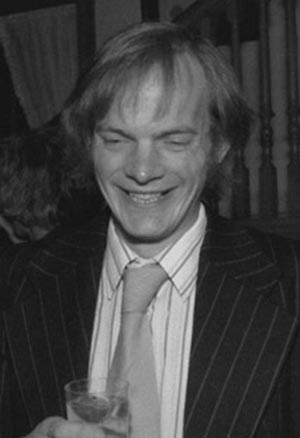
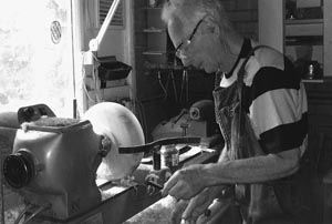
David Lane, ca. 1980.
(Photograph courtesy of Ashley Dunn.)
David’s PhD was at University College London with Avrion Mitchison, one of the last great eccentrics of British science. The son of the novelist Naomi Mitchison and her politician husband Dick, Av came from the fine tradition of scientists who do what they do for the fun of it and don’t give a hoot for convention. Unsurprisingly, he was good friends with Jim Watson, who was fascinated by Av’s mother, lifestyle, and personality, and Watson was his best man when he married.
Av’s style in the lab was, to quote David, “wonderful and awful at the same time.” The Tumour Immunology Unit, which Av ran, was funded by ICRF and was one of the hot places to be in the immunology world at that time. In contrast to the rest of UCL, which was very old and fussy, Av’s unit was painted in bright colours and was full of bright people to match the décor. However, even Av admits that David was neglected when he started work there, partly because Av was preoccupied with the demands of running a new unit, but partly because that was just what Av did—you were given space, equipment, and a pool of clever people to talk to, and then left to sink or swim. David spent his first year virtually unsupervised, trying to get a horribly difficult assay to work, in the meantime watching the other students and postdocs knocking out Nature papers. He was unsurprised, if upset, when Av told him he thought he was no good and should give up, but because it transpired that Av was always firing graduate students and then unfiring them, David survived. He was then plunged back into gloom by the news that Av had hired an American postdoc, Don Silver, to work on exactly the same project, and this time was only prevented from quitting by the fact that he had acquired a new girlfriend, Birgit Muldal. The love of a good woman (whom he ended up marrying in 1975) was enough to keep him in high enough spirits to continue, and it was just as well, because Don, rather than being a competitor, was extremely helpful, ending up as David’s de facto PhD supervisor.
Between them, Don and David made a very good team. In between endless conversations regarding life and science, Don taught David to think critically, and also persuaded him to work a little harder (David claims, slightly improbably, to be intrinsically lazy). Don had a background in genetics, and with this added bonus, the pair started to make great progress, publishing a paper in the Journal of Experimental Medicine, then the top of the tree as far as immunology was concerned, in 1975. Av was delighted, promoted David to star student status (he now recalls him as “the best student I ever had in University College”) (Mitchison Web of Stories), and fixed him up with a postdoctoral position with Bill Paul, a top U.S. immunologist working at the National Institutes of Health (NIH) in Bethesda, Maryland. In passing, Av also sorted out the newlywed Birgit and David’s housing issues by installing them in the flat at the top of his rambling old house in Islington, a typically generous gesture. Birgit was finishing up a postdoc at Imperial College and also had a job offer at the NIH, so David just needed to find something to do for the next nine months, and they would then be in sync. All seemed set for a satisfactory transatlantic transfer from one immunology guru to another, followed by a long and happy immunological future.
It was not to be; the world beyond immunology thrust its way into David’s life, and neither David nor Birgit ever made it to Bethesda. In David’s words, “something happened then that was really strange—rumours got around that I knew how to iodinate proteins. I’d been making my pure protein by setting up a radioimmunoassay for it which was very important for the quantitative immunology we were doing. It was right on the edge—very difficult to do, but it just about worked.”
Being able to successfully radioiodinate proteins was, as David indicates, a big deal in those days, and was also a really good way of accidentally dosing yourself with a fair whack of potentially toxic radiation. David’s predecessor in Av’s lab had, in fact, messed up to such an the extent that he was the Health and Safety Department’s poster boy for the “how not to do radioiodination” safety material. The procedure is a way of labeling proteins by attaching hot 125I to them and is very useful for monitoring molecules present at very low concentrations such as the purified protein David was using in his radioimmunoassays; 125I emits γ-rays, so that it is easily detected even when present in tiny amounts. The flip side of this convenience is that γ-rays are so energetic that they are very good at killing cells, which is good if you have cancer, where they are used in radiotherapy, but bad in normal life, where they can not only destroy healthy cells but also mutate them. 125I can be absorbed quite easily whilst doing experiments, mostly because the labeling protocol calls for the radioactive iodine to be produced from sodium iodide and reacted with the protein recipient; the free 125I produced during the reaction is highly volatile and therefore very easy to inhale. Once in the body, 125I migrates to the thyroid gland, where it can profoundly damage the tissue. Av’s unit did at least have a fume hood, which gave some degree of protection, but even so, trying not to breathe too much was the preferred option in the crucial parts of the experiment.
The rumours of David’s interesting skill set reached Lionel Crawford at the ICRF via Mike Fried’s wife, Nancy Hogg, one of David’s fellow inmates at the Tumour Immunology Unit. Lionel, as related previously, had realised that the antisera for detecting SV40 T antigens were awful, the assays were, in his words, “cumbersome, irreproducible, and everything else,” and that without improving them, nobody was ever going to be able to work out what was going on. Rather than struggling on with what he had, Lionel had had an idea regarding how to quantitate antisera properly but needed a competent immunochemist who wasn’t scared of radioiodination to convert his theory into practice. Now, it seemed, that person might exist. He contacted David and asked him to come over to the ICRF for a chat.
Lionel, meticulously well organised both in his thinking and his experimental work, was about as different from the wildly eccentric Av as it was possible to get, but the difference in style was intriguing, the project was interesting, and the salary was seriously good compared with David’s PhD stipend of just under £500 a year (equivalent to £4000 in current money). David signed on, two years and nine months into his PhD, without having written up his thesis, which would have to be worked on in his spare time (or more realistically, because he didn’t have any spare time, when he should have been sleeping). Lionel was delighted—he had managed to hire someone who was confident with radioimmunoassays, confident with immunising animals, good at the bench, clever, keen, and more than happy to work independently—postdoctoral gold dust.
The ICRF was virtually unknown territory to David and came as a shock to his system. Life in the Tumour Immunology Unit had the speed and ethos of an arthouse movie, with long theoretical discussions during endless coffee breaks, where one’s intellectual prowess was perhaps more valued than practical ability. By contrast, the ICRF Fifth Floor was more Raiders of the Lost Ark crossed with a hefty dose of Jaws. Work was fast, precise, and biochemical, with exciting new data seeming to appear on an almost daily basis, and the predatory sharks cruising the floor did not so much discuss ideas as violently shred them, biting chunks out of their opponents’ egos in the process.
It didn’t take David long to realise that the inhabitants of this brash new world, although doing great things in many areas, had an immunochemical blind spot. Strangely, because they were rigorously quantitative when it came to the molecular biology techniques that they were pioneering, for immunoprecipitations they were using a bunch of unquantitated antisera made from assorted rodents, with little idea of how good the antisera were. This was all very well when everyone thought they were just looking for one 100-kDa protein, because if that protein showed up, the responsible antiserum was judged a success. Hunting for new SV40 T antigens was a different matter, however; how could you tell whether the many extra bands that always appeared along with the 100-kDa band were specific, or just background? You could get some of the way by comparing immunoprecipitations from tumour-bearing animals with those from tumour-free animals, but even then, the gels run to separate the immunoprecipitated proteins still had so many dodgy-looking regions that assigning one band to one protein was impossible. To quote Lionel: “Once you see things further down the gel, you think ‘ooh, what’s that?’ And the answer is, ‘it’s a whole mess.’ ” Alan Smith’s lab was trying to get around this problem by translating SV40 proteins in cell-free extracts to get rid of some of the background, but even they hadn’t got very far.
The reasons for the awful data were twofold: the crude way in which the antisera were made, and the subsequent immunoprecipitation assay. In those days, antiserum was simply whatever was left over after the blood cells and clotting factors were removed from blood; the resulting clear, yellowish liquid contained a mixed bag of whatever antibodies happened to be circulating at the time of the bleed. Therefore, although it was likely that an animal with SV40-induced tumours would have a lot of SV40-specific antibodies, what the mix of antibodies saw in the tumour and how well they worked to bind antigens could not be guaranteed. As a result, different batches differed wildly in efficacy. Further problems crept in during immunoprecipitation; the antiserum was mixed with a radiolabeled cellular protein extract, and then the small number of antibody–antigen complexes that had formed had to be pulled out from the uninteresting stuff by a variety of methods, all of which were long-winded and inefficient. As a final hurdle, the huge number of other proteins knocking around in the labeled cell extract meant that some of the stickier ones always sneaked through to the end to produce nonspecific background; it was almost impossible to get rid of them by washing the immunoprecipitates because thorough washing tended to destroy the genuine antibody–antigen complexes too.
In the mid-1970s, a new immunoprecipitation protocol appeared that rapidly eclipsed the other methods. Protein A is a surface protein made by the bacterium Staphylococcus aureus as part of its defence against being killed when it infects a new host. In its role as point man for S. aureus, protein A binds tightly to many antibodies and so traumatizes the cells carrying them so that they either kill themselves or stick their fingers in their metaphorical ears and shout “la la la” whenever they see any new S. aureus heading their way (in immunospeak, protein A, a superantigen, elicits apoptosis and tolerance induction).
Because of its ability to bind antibodies, researchers realised that using protein A would be a great way of pulling down immunoprecipitated proteins. Either bound to heat-killed S. aureus or subsequently to little beads rather like miniature frog spawn, protein A rapidly attaches itself to antibody–antigen complexes with a tenacity that allows vigorous washing to be deployed to eliminate nonspecific proteins. The results are far cleaner, and the experiments quicker and less fiddly. In David’s words: “you just squirt everything in and it works beautifully.”
David’s first tasks in Lionel’s lab were to find a way of testing all of the existing antisera and then to devise a new method of making more specific reagents for the future. Both objectives required him to look at the antibody–antigen complexes formed in immunoprecipitations from a different viewpoint, asking questions regarding the antisera rather than the antigens. To do this, he and Lionel had come up with two rather good ideas: to use radioiodinated protein A as a marker to detect how much complex was present, and to trap and then count the immune complexes on glass fibre filters.
Lionel and David published their paper, “An Immune Complex Assay for SV40 T Antigen,” in January 1977, a few months after David’s arrival. Their new assay started off as a normal immunoprecipitation, mixing antisera with cell extracts made from one particular line of SV40-transformed cells. Then, instead of spinning them down, the samples were put onto glass fibre filters, which trap immune complexes but not other proteins, and washed very carefully. Protein A labeled by David with 125I was added to the filters, and after some more washing, the amount of radioactivity left on the filters was measured. The better the antibody bound the T antigens in the cell extracts, the more complexes stuck to the filters, and the more 125I was left. The new quantitative tool allowed David to test all of the lab’s antisera, bin all the terrible ones, and rank the remainder according to their strength of binding to tumour antigens.
David had achieved his first objective very quickly, and he rapidly realised that it had also given him a way of completing his second task, to make better reagents. However, Lionel was not around as a source of help and advice for this next stage; even before their paper had appeared, he had gone off on a year’s sabbatical to Stanford, to work with George Stark, a great mate of his. Fortunately, this event, which would have struck fear into the hearts of many novice postdocs, was so similar to the benevolent neglect David had encountered as a PhD student that it didn’t bother him at all: “I didn’t expect in any way to be supervised, which is amazing when you think about it now.”
Slightly more alarming than being abandoned by his supervisor were the territorial battles after Lionel’s departure. Alan Smith and Bob Kamen were both given permanent jobs as proper lab heads whilst Lionel was away, but the jobs didn’t come with more space. Possession being nine-tenths of the law, their territorial ambitions meant that David and Peter Piper, Lionel’s other remaining postdoc, had to mount a vigorous defence of Lionel’s office space and benches. At one point, David himself became the subject of a takeover attempt from the Smith lab, but when Alan came to him with a list of experiments he should perform that week, David just told him that he wasn’t interested and carried on with his own projects. Eventually, things settled down, and everyone got back to what really mattered, the research. Joint lab meetings and seminars carried on, and the big open-plan Smith/Kamen/Crawford lab contained some amazingly good people, who were all happy to talk to David: “I had Bob [Kamen] and Richard [Treisman] and Rich Condit on one side, and on the other side was Eva [Paucha] and Alan [Smith] and a vast team including Tony Pawson and Frank McCormick.”
Lionel left David a valuable bequest before heading off to California; his technician Alan Robbins, who was very good, very experienced, and as a bonus, was a lot of fun. David and Alan got on famously: “We just treated each other as equals and worked together really well. It was just one of those systems where in the morning you’d do something and in the afternoon the other person would finish it. You knew you could completely trust the other person. The only time it broke down was Friday afternoon. We literally used to run to the pub at 5pm on Friday—it was hilarious.”
At the end of David and Lionel’s paper on quantitating SV40 antisera, there is a beautiful photo of an immunoprecipitation run out on a gel. It is squeaky clean—there are hardly any bands to be seen in the track precipitated using anti SV40 antisera—and, most importantly for David’s next step, there was an awful lot of one particular band running two-thirds of the way up the gel: large T antigen. David, in his reading of the immunology literature, had seen a paper in which proteins could be cut out of gels and used to immunise animals to make antibodies, and he realised that if he did the same with large T, he could make antiserum specifically directed against it. This was a big step forward, because all of the previous antisera had been made against whole tumour cells. Having a T-specific antiserum would be like the difference between knowing a postcode and knowing a house number within that postcode. You could go straight to the front door you wanted without bothering the neighbours.
Leaving aside its use as a great immunogen, pure T antigen would also be an invaluable asset for getting going with biochemistry and the new art of protein sequencing. People could study what it was doing and also find the amino acid sequence of its front and back ends; turned back into the DNA three-letter code, the sequence would localise the beginning and end of the large T gene on the viral genome and also match it with the mRNA from which it was made. In retrospect, it isn’t surprising that Alan Smith had been keen to suborn David, because this was exactly what his lab wanted to do. The difference, however, was that they were making small amounts of large T in test tubes and would have dearly loved to get their hands on the cellular protein purified by David’s technique.
David and Alan Robbins got in a big order of monkey kidney cells from Kit Osborn, Lionel’s cell culture technician, infected them with SV40 virus, and made a lot of cell extract. After a massively scaled-up immunoprecipitation, they ran the result out on a big gel, cut out the large T band, solubilised it, and then headed off to the animal house with their precious immunogen.
The story so far, but for the cameo appearances of Kit, Birgit Lane, and Nancy Hogg, has been sadly lacking in female interest. In human terms, this will unfortunately continue, because the Fifth Floor was a very male environment, perhaps accounting for some of the testosterone-fueled antler clashing and similarly daft behaviour that went on there. In the rabbit world, however, a heroine enters at this point; large and white, with big floppy ears and a benevolent expression, Lucy the antibody rabbit single-handedly (-pawedly?) provided David and Alan with the new antiserum they needed. After injection of the large T solution, she produced vast amounts of really good anti-T antiserum whilst remaining in excellent health, unlike some of the other rabbits in her room, who perished in an epidemic of Snuffles, a nasty bunny illness that does exactly what it says on the box. The Snuffles epidemic nearly put paid to the experiment, because the animal technicians wanted to clean out the affected room by culling poor Lucy; fortunately, they relented, meaning that a daring plan to smuggle Lucy out and keep her at home was never executed. Lucy went on to live a productive and reasonably comfortable life (but for the need for regular injections) and produced enough fantastically good antiserum for several years of experiments. She is still remembered with great respect.
David and Alan wrote up their results and submitted a paper in late 1977 presenting their new antiserum. The paper showed that Lucy’s anti-T antiserum was incredibly clean and very potent, five times as active as the best hamster antitumour antiserum. The relative size of hamsters and rabbits also meant that she had produced the same amount of antiserum as 500 tumour-bearing hamsters, a considerable saving on all fronts. Most interestingly of all, with the background of nonspecific proteins reduced to almost nothing by David’s technical expertise, it was clear that the anti-T antiserum saw not just the large T antigen with which Lucy had been immunised, but also two smaller bands. One of these, the really small one, was SV40 small T, which Lionel, still in Stanford and collaborating with Paul Berg’s lab, had recently shown was a splice variant of large T; it made sense that the antiserum should recognise this too because large and small T have a fair bit of common sequence. The other band ran at ∼60 kDa; it was not clear what it could be, and David and Alan, instead of speculating, just left it at that, merely saying in the Discussion to the paper that “its relationship to large T and small T is currently under investigation” (Lane and Robbins 1978).
The dry scientific language disguises the fact that before David and Alan’s paper, that position on a T antigen immunoprecipitation gel had always corresponded to the awfullest, messiest, region. This was the first time that a single protein had emerged from the confusion. Importantly, because it had been immunoprecipitated with anti-T antiserum, it had to have some relationship with SV40 large T and wasn’t just a very persistent background band of no interest. Therefore, the “currently under investigation” of David and Alan’s discussion of the 60-kDa band translates as “We are busting a gut to figure out what it is before somebody else does.” David was well aware that he would have to really crack on to beat the opposition when it came to identifying it.
In addition to the lab-based excitement, there was a lot else going on in David’s life at that time. For a start, he was desperately trying to write up his PhD thesis for submission. In those days, you typed your thesis by hand, referenced it by hand, and made the figures yourself, and David and Birgit spent a lot of nights taking turns to type until their hands hurt in their new house in Chiswick, where they’d recently moved. The other more startling event was that David was now in possession of a rather good extramural job. Av, keeping a fatherly eye on him, had phoned David up one day and suggested that he applied for a lectureship in immunology at Imperial College. Following an interview, David had duly been appointed, despite lacking a doctorate and being only 25. As mentioned before, David claims it was mostly luck, not talent: “I wasn’t extraordinary—it was just a set of circumstances and they wanted someone very cheap! At the interview they said, ‘We can afford a Rover but not a Rolls Royce’ in terms of equipment, so they bought me a couple of things and I set up a little lab there, continuing some of the things from my PhD.” It was just as well that David had acquired a motorbike, because after his appointment, he spent an awful lot of time commuting between home, Imperial, and the ICRF (a juggling act that pales into insignificance against his subsequent multijob existence in Singapore, Dundee, and London).
Matters didn’t stop there, however, because yet another job offer materialised, once more via Mike Fried, who was inadvertently evolving into David’s scientific Fairy Godfather. Having first brought him to Lionel’s attention after Nancy Hogg’s tipoff, Mike now introduced David to Joe Sambrook from Cold Spring Harbor, who was over on one of his regular romantic/scientific visits to the ICRF. Sambrook liked the look of David and asked him to come to Cold Spring Harbor as a postdoc, to work on hybrid SV40/adenoviruses. Imperial, who had been very relaxed about David spending most of his time at ICRF, seemed equally happy for David to go to America, as long as he showed up to give his lectures and they didn’t have to pay him. Birgit got an offer from another lab at Cold Spring Harbor, abandoning her NIH plans, and everything was set, with a start date of late 1978. ICRF did very well out of these arrangements: “I was at ICRF for two years in the lab, but as an employee, probably nine months. I was a very cheap deal for everybody, really!”
Lionel’s return from California must have been a bit unpleasant for him. He had spent a happy year doing what he loved best—experiments—and had ended up with some great papers to show for it; as mentioned in the previous chapter, his discovery of how SV40 small T was made was the first ever demonstration of splicing in a coding region. He returned to find that his two postdocs, Bob Kamen and Alan Smith, had not only been transformed into independent lab heads but had been given some of his technicians, and that his own lab had consequently shrunk to almost nothing. Bob remembers him being remarkably generous about it: “Alan and Lionel and I got together and I thought it would be controversial but Lionel said ‘Fine, let’s split it three ways.’ So I got the left two bays, Lionel got the middle two bays and Alan got the right two bays. It was hard to tell if he minded—he was very, very British, non-confrontational. I don’t think he minded. I was certainly very happy he hadn’t created a problem.”
David had also changed: “When Lionel came back, he met a very different creature. He met someone who’d got an independent position and written a rather nice paper in a journal and had discovered this other protein, and hadn’t put his [Lionel’s] name on anything! I didn’t know, because Av hadn’t taught me, how things worked in the rest of the world. The work with Alan, I just told Lionel the paper had been accepted by Virology, and wasn’t that good? When he said he was surprised I hadn’t put his name on the paper, I was truly astonished! Looking back, I must have seemed very arrogant, but it was more naïvete than anything.”
Leaving aside the charged personal situation, both Lionel and David were very excited by David’s mystery 60-kDa protein. In June 1978, David got his PhD, finally justifying the “Dr. David Lane” sign on his Imperial office door, and was able to really turn his mind to what could be going on.
In the first half of 1978, David had spent a lot of time tinkering with the fine detail of his immunoprecipitations, as had Lionel, who, somewhat to David’s chagrin, was mirroring the work to get the gels looking better. There were several items that could be tuned up, and all were attended to. David realised that preparing the cell extracts by gently popping the cells open with a smidge of detergent rather than the accepted method of plunging them into dry ice repeatedly meant that the proteins inside didn’t fall apart. Intact protein meant fewer bands on gels, and more material running at the right size, and was especially important for large T, because it had a nasty tendency to disintegrate into a set of bands running around 60 kDa, confusing any analysis of that region. The antiserum, now that it had been properly tested for activity, could be used in minimal amounts (1 µL—a thousandth of a millilitre—of Lucy’s antiserum was enough to get a good clean immunoprecipitation), and pulling antibody–antigen complexes from the extracts using protein A resulted in even more reduction of background.
Having optimised the methodology, David then started comparing cell lines for amounts of 60-kDa protein. As he’d observed previously, untransformed cells had no detectable 60-kDa protein at all, and, in fact, the cells in which he’d first seen it, the SV40-infected monkey kidney cells, didn’t contain very much either. Far better was an SV40-transformed line, SVA31E7, which had boatloads of it. With more protein visible, it was also easier to estimate size; comparison with a good marker ladder (a mix of proteins of known molecular weights, run beside samples) showed that a more accurate weight estimate for the mystery protein was 53 kDa. It was duly christened 53K, which was a bit vague, but would do until someone worked out what it did and renamed it accordingly.
The big question with the 53K protein pertained to its relationship with large T. Was it showing up on the immunoprecipitations because the relationship was familial, as for small T? Both the large T and 53K bands were always there following immunoprecipitation by lots of different antisera, either Lucy’s rabbit anti-T or the old antitumour stuff, and each protein was equally well recognised, but this didn’t necessarily mean that they were related. The killer experiment would be to do an immunoprecipitation, run the samples on a gel, cut out the large T and 53K bands, and then see whether the isolated bands could still be recognised by Lucy’s anti-T serum in an 125I filter-binding assay.
David did a spot of breath-holding and involuntary self-iodination to make some more radiolabeled protein A, and the experiment went ahead. It had a very unexpected result; the isolated large T band was happily recognised by Lucy’s antiserum, but the 53K protein was not seen at all, despite numerous attempts to prove the contrary. The conclusion had to be that the 53K protein did not have any of the same antigenic determinants as large T and wasn’t related to it at all. So what on earth was it?
The most obvious answer, that it was an artefact and the 53K protein was just sticky rubbish, could be quickly excluded in David and Lionel’s minds; they knew that their extracts and gels were irreproachably good. This left only one possibility, that the relationship between large T and the 53K protein was not familial, but conjugal; the 53K protein, like an overpossessive spouse, was so clingy that in anti-T immunoprecipitations it always came along for the ride. In fact, it was bound so tightly that only separation on a gel could tear it away.
So far, so good, but this still didn’t solve the question of where the 53K protein was coming from. Because it only showed up in SV40-transformed cells, logic dictated that it ought to be made by the SV40 early region, but this was becoming increasingly unlikely; that year, the SV40 DNA sequence was coming out in dribs and drabs, and it showed that there was simply no room in the viral early region for another completely different protein in addition to large and small T. Something else, something novel, had to be going on.
Lionel and David suddenly realised that what they were looking at might be partial vindication of the years of pouring millions of pounds and dollars into research on small DNA tumour viruses. The whole point had been to use the viruses as a way of finding the cellular control mechanisms they were subverting to cause cancer, and it followed that to subvert the control mechanisms, contact had to be made with the cellular control proteins. That was it! The 53K protein must be cellular, not viral. Large T, far from establishing a conjugal relationship with it, had forced the transformed cell to switch the 53K protein on and was holding it hostage.
Thomas Edison famously said that genius was 1% inspiration and 99% perspiration. Whatever the ratio for David and Lionel in the matter of the 53K protein, once they realised that large T was contacting the cell via the 53K protein, it was as if one of Edison’s patent incandescent light bulbs had come on in their heads. An interaction that tight had to be important for transformation, although how, exactly, was not a question they would be able to answer any time soon. What was most pressing was to make the case for the cellular origin of the 53K protein as watertight as possible, and publish. Their technical expertise meant that they had an edge over the competition, but they couldn’t rely on that forever.
First up was to test all the other antitumour antisera in the filter binding assay, to see if they behaved similarly to Lucy’s anti-T serum. Surprisingly, although most of the antisera were identical in their inability to recognise the gel-isolated 53K protein, one antitumour serum bucked the trend, still seeing both proteins after gel purification. The conclusion was that in that particular tumour, the mouse host had made antibodies not only to T antigen proteins, but also to the cellular 53K protein. This had to be properly proven, so David did a collaborative experiment with Eva Paucha, Alan Smith’s postdoc (extremely nice, very talented, and very beautiful, Eva Paucha was Alan’s partner not only in the lab, but outside it; her early death a decade later at the age of 38 was a huge tragedy). Using the Smith lab’s cell-free translation system, Eva showed that there were conditions under which the 53K protein could be detected by the oddball antimouse serum even when it wasn’t complexed to large T.
Having the strange mouse antiserum allowed David and Lionel to do one final very important experiment. If, as they thought, the 53K protein was important for transformation and cancer, then it should show up not just in SV40-induced tumours, but in other sorts too. David did one final filter binding assay, using protein extract made from polyoma-transformed cells, which had never had so much as a sniff of SV40. All of the anti-SV40 antisera failed to bind anything, except for the strange antiserum, which saw a protein near enough in size to the 53K protein to suggest that it was very closely related, if not the same item.
David and Lionel wrote up their data in a short, three-figure paper entitled, “T Antigen Is Bound to a Host Protein in SV40-Transformed Cells,” sent it to Nature in November 1978, and sat back, fingers crossed. If the reviewers liked it, it would with any luck be published before the turn of the year, thus forestalling any arguments about who had got there first. Sadly, the reviewers did not like it at all; it came back with all sorts of objections attached to it, most of which were also voiced by other members of the SV40 community when Lionel sent them preprint copies (the generosity of those days, when even big results were shared with other labs before publication rather than hugged to one’s chest for as long as possible, is astonishing to the modern eye). The main complaints seemed to be that (1) nobody believed it; (2) despite that, everyone had done it already; and (more coherently) (3) there was no proof that a complex had formed other than the immunoprecipitation data, and something else was needed. Because the last point was something tangible that David and Lionel could address, they did a hasty biophysics experiment that brought the number of figures in the paper up to four. David’s opinion of this “reviewers’ experiment” is unflattering to all concerned: “The critical thing that we did that satisfied them, although it was a crap experiment, was to run some gel filtration columns and show that they [large T and the 53K protein] came out together. But if you look at the figure, it’s pretty awful!” It was enough, however, and Nature accepted the paper at the end of January 1979.
By this time, David had moved to Joe Sambrook’s lab at Cold Spring Harbor; thus, the final revisions were completed by transatlantic phone call and the proofs corrected on a flying visit home, in Lionel’s house in Highgate. The paper came out on 15 March. Unfortunately, by this time, the competition had caught up, and so officially, David and Lionel published the discovery of the 53K protein in the same year as three other groups, rather than the year previously.
The speed with which the other labs got their papers out is very illuminating. As had happened to Bob Kamen during the discovery of splicing, other people had got similar stuff but hadn’t hit on the right explanation for what was going on (gallingly for the authors involved, three papers published in 1978 identified a band corresponding to the 53K protein but failed to realise its significance). As Lionel says, “so many of these things, you just think ‘ah, yes!’ Several people had the data and once they were provided with an explanation were able to write up very quickly.”
Two more papers, by Daniel Linzer and Arnie Levine at Princeton, and from Pierre May’s group in Villejuif, also looked at the 53K protein from the SV40 angle and confirmed David and Lionel’s data. Linzer and Levine additionally found a protein that was similar or identical to the 53K protein in embryonal carcinoma cells. This result was important because it showed very clearly that the 53K protein was present in a cancerous cell type that had nothing to do with SV40 or polyoma, implying that it might have a more general role during transformation. May’s group published more of a roundup of all of their immunoprecipitation data, with no speculation as to what might be going on beyond the observation that some of the bands were potentially of cellular origin. What their paper does illustrate very well is the sheer number of bands that it is possible to immunoprecipitate with anti-SV40 tumour antisera—it is small wonder that the authors were unable to work out which might be of greatest importance.
The other paper coming out that year, from Albert DeLeo and Lloyd Old from the Memorial Sloan-Kettering Cancer Center in New York (together with colleagues from the National Cancer Institute in Bethesda), was crucial in establishing the wider relevance of the 53K protein, showing that Linzer and Levine’s embryonal carcinoma data were not just a one-off. DeLeo’s paper provided the link to other forms of cancer and showed the protein was not just a virally induced phenomenon. In it, the investigators identified a tumour antigen of cellular origin that was never present in normal cells but always showed up in transformed cells, irrespective of the carcinogenic insult. Every transformant they tested, whether spontaneous or chemically or virally induced, contained the antigen, which they called p53.

Their competitors’ interest in David and Lionel’s data was not mirrored with much enthusiasm by the wider DNA tumour virus field. Even Fifth Floor reactions to the paper were muted. The Smith lab published a beautiful Cell paper subsequently in 1979 that established conclusively that the 53K protein was of cellular origin and had a completely different protein sequence from that of large T, and they continued working on the problem after Alan’s departure for the National Institute for Medical Research, but in the polyoma-oriented labs, the reaction ranged from Not Interesting: “I was anti—I just thought it was another host binding protein” (Mike Fried); “I didn’t really focus on it” (Bob Kamen), to Possibly Interesting: “Because it was a band that wouldn’t go away when subject to real stringency, there was a sense that this might be something” (Adrian Hayday). Outside the ICRF, in his new home of Cold Spring Harbor, David encountered outright hostility. “[One of the senior staff investigators] came up to me and said they didn’t understand how I could publish something like that, as it was obviously wrong. I had a complete meltdown! People didn’t accept the immunological idea—they thought that for a protein to bind that tightly so that it could resist washing, processing and all that, was just not possible.” David, with his immunological background, knew that there was established precedent for such tight protein–protein interactions in the immune system, but sadly, his less well-informed critics remained unconvinced.
Immunological ignorance aside, the main reason for the underwhelming response to the 53K protein was that by the late 1970s, DNA tumour viruses were widely regarded as the best tools for dissecting the mechanisms of eukaryotic transcription, translation, and DNA replication, but had fallen out of fashion in terms of cancer research. The big hitters in the molecular biology of cancer were now the RNA tumour viruses, thanks to Harold Varmus, Dominique Stehelin, and Mike Bishop’s 1976 discovery of the nature of retroviral oncogenes.
In a paper that won Varmus and Bishop the 1989 Nobel Prize for Physiology or Medicine, the v-src oncogene of Rous sarcoma virus was unmasked as a cellular gene gone bad; Rous sarcoma virus had picked up a copy of an important cellular control gene, c-src, and mutated it into a form that caused unregulated cell proliferation, and hence cancer. Researchers everywhere leapt enthusiastically onto the retroviral oncogene bandwagon, working on what c-src and v-src might be doing, and also trying to identify further captured cellular oncogenes in their own pet retroviruses (e.g., at the ICRF, Tom Curran, a PhD student with Natalie Teich, found the v-fos oncogene in the FBJ murine osteosarcoma virus). This flurry of activity rapidly confirmed that the capture and corruption of cellular genes, so-called proto-oncogenes, had happened multiple times, and that the genes in question played important roles in both normal and cancerous development.
The balance tipped even further in favour of RNA tumour virus research in 1981, when Bill Hayward, at the Rockefeller Institute in New York, showed how Avian leukosis virus, ALV, causes cancer. ALV belongs to a second group of retroviruses that do not carry their own personal oncogene, but are still extremely tumourigenic because of a peculiarity of the retroviral lifestyle; after infecting a cell, all retroviruses make a DNA “provirus” copy of themselves using reverse transcriptase and then insert the provirus into the host cell’s DNA, where it becomes a passenger in the genome. When Hayward examined where in the genome his ALV provirus had put itself, he found it next door to a growth-controlling gene called c-myc, which had already been identified as the cellular proto-oncogene kidnapped by another virus, MC29. Hayward showed that powerful transcriptional control elements in the ALV provirus were forcing the cellular c-myc gene to make inappropriately high amounts of c-Myc protein, driving cells into constant proliferation and causing a tumour to develop. He christened this new method of tumourigenesis “insertional mutagenesis.”
RNA tumour virus research really seemed to have done its job and cracked the cancer problem; with the retroviruses, Mother Nature handed researchers the means of identifying scores of cellular genes important for regulating growth. Although it would not be trivial to work out how these genes worked, it was as though the doors had suddenly been opened into a hitherto locked building, and light was shining into many unexplored rooms.
In this new dawn, the DNA tumour viruses began to look almost irrelevant. Their oncogenes did not have cellular counterparts. They did not cause insertional mutagenesis—they seemed to integrate randomly in the genome. The 53K protein was one of only a few established links to the cell, and it was, frankly, a boring band on a gel, with nothing to recommend it other than its insistence in showing up in a lot of transformed cell lines. Figuring out what it did would require it to be cloned, and cloning cellular genes was incredibly hard. Far better to work on retroviruses, with their dinky little genomes that made cloning individual genes comparatively easy.
Research on the 53K protein did continue, but only in the small number of labs with an existing interest; new recruits were very thin on the ground. The smallness of the field did mean that one problem dogging the protein could be resolved relatively harmoniously—the question of what to call it. Everyone who had published on it had come up with a different name—the 53K protein, Non Viral T Antigen, the 54K protein, p53, phosphoprotein53. After some arguing, the field agreed on “p53,” and thus it has appeared in all subsequent literature. Ironically, p53 is not 53 kDa in size; the protein has some oddities that make it run abnormally slowly on gels, and its weight is actually ∼43 kDa or less. However, by the time people figured this out, the name had stuck.
The early work on p53 had a dual focus. Firstly, most labs harboured at least one unfortunate soul stretching the existing technology and their own ingenuity in the race to clone the p53 gene, a feat accomplished in 1982 by Peter Chumakov and colleagues in Moscow, closely followed by Moshe Oren in the Levine lab, and Lionel’s group. Secondly, efforts to characterise the p53 protein were ramped up, made easier by great improvements in the antibodies against SV40 large T and the 53K protein. A new, powerful immunochemistry reagent, the monoclonal antibody, had appeared in 1975, when George Köhler and César Milstein at the Laboratory of Molecular Biology in Cambridge fused antibody-producing B cells and myeloma cells to make hybridoma cell lines that grew in culture indefinitely and made just one antibody type, directed against a single antigen on a single protein. To resume the postcode analogy of antibody binding, if antitumour antisera see a postcode, and anti-T antisera a specific house within that postcode, a monoclonal antibody can go into the house and find the toaster in the kitchen. The ability to achieve such exquisite specificity in antigen recognition has revolutionised both experimental science and medicine, where monoclonal antibodies are widely used as “magic bullet”–style therapeutics. Not surprisingly, their invention garnered Köhler and Milstein a Nobel Prize, in 1984.
David, at Cold Spring Harbor, and Ed Harlow, a new PhD student in Lionel’s lab at the ICRF, made some of the earliest and best monoclonals against SV40 large T and p53. David was having an interesting time in Sambrook’s lab: “When I went to ICRF I thought I’d landed on Mars, but when I went to Cold Spring Harbor it was definitely Mars! Joe Sambrook was very ruthless. He had two types of postdocs, regular American ones, who were properly selected and carefully interviewed and came in to work on particular projects, and then he had a bunch of bright English people he threw in there. If you weren’t any good you just left. There was no equipment. There was nothing, so people would steal. It was chaos—disorder, total selfishness and absolute obsession. It’s kind of miraculous what we all managed to do there.”
David and Ed Harlow had overlapped briefly in Lionel’s lab and hit it off instantly. This was unsurprising because, to be honest, Ed’s brand of laid-back friendliness is hard for anyone to resist. Born in Southern California and ending up in Tulsa, Oklahoma after a peripatetic childhood, Ed decided he wanted to see the world and wrote to Lionel for a job. He pumped gas for six months to make enough money to come and arrived entirely grantless, clad in Oshkosh dungarees, and equipped with nothing but intelligence, ambition, and a willingness to work (and party) very hard. Lionel, entirely charmed, found him the money to stay for a PhD. It quickly became evident that beneath his happy-go-lucky exterior, Ed was the real deal: a thinking, original scientist who was also incredibly good at the bench. His project consisted of making monoclonals and plugging them into the SV40 system, and he published eight papers during his three years at the ICRF. With Frank McCormick, a postdoc with Alan Smith and yet another scientific star in the making (a pioneer of the biochemical dissection of cancer and of targeted cancer therapy, Frank became Director of the UCSF Cancer Center in San Francisco), Ed did the reverse experiment to Lane and Crawford, showing that a monoclonal antibody against p53 would coprecipitate large T antigen, and getting himself a Nature paper in the process.
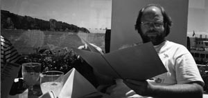
Ed Harlow orders lunch, ca. 1992.
(Photograph courtesy of author.)
Now a Harvard Professor of many years’ standing, and a Senior Advisor to Harold Varmus, Director of the U.S. National Cancer Institute (and an old friend from his ICRF days), Ed’s memories of the ICRF are what everyone should get from a good PhD experience: he had a great time, found a lot of friends, and began to realise he could make it as a scientist:
ICRF was a breeding ground for a large number of folks that have gone on to do exceedingly well. I don’t understand what made this group special, other than the good science at ICRF attracting committed individuals who did well over time. One thing that might be true is that we did get pushed and subsequently learned that we could do useful things. That confidence was built at least in part from the environment. In a productive but somewhat perverted way, the ICRF 5th floor lab heads were great models. Their allegiances to and disputes with one another changed over time, but their commitment to science was always first. I learned that one didn’t have to be deep personal friends to respect the quality of science that was being produced. [However], I am now very committed to not doing science with folks I don’t like! A simple rule that I have adhered to for years is that if I don’t want to have a drink with someone, I don’t want to work with them.
In many ways, ICRF and London were a rite of passage—I continue to view London as the most important stage of my career. I came from a spirited but second-class training, and I learned what world class science was at ICRF: important questions, rigorous answers, and the excitement of discovery. I learned that I thrived in big cities, that having access to first class cultural events—particularly art and theatre—was important to me, that I wanted to work in the best scientific environments, and that I could compete at that level. The scientists from my ICRF days have become an important circle of friends whom I continue to cherish. I would go to huge lengths to help them if they needed it, and I’d be surprised if they wouldn’t reciprocate if I called on them for any type of favour.
Ed’s evident star quality made him an inevitable target for recruitment to Cold Spring Harbor, and he went off there in 1983 to work on the adenovirus E1A oncogene, leaving behind a case of Dubonnet for Kit Osborn in thanks for her tissue culture virtuosity. No one was more pleased for him than David: “Ed’s an amazing guy, a very generous person and easy to get along with. He was very excited about monoclonal antibodies and he got more and made better ones than me, although it was OK as we were very friendly and we swapped the reagents around a lot. Cold Spring Harbor realised they needed somebody who could make monoclonal antibodies and I recommended him for the job. It worked out incredibly well.” Despite David having left Cold Spring Harbor to finally take up his lectureship at Imperial College by the time Ed arrived, the two men stayed close, ending up coauthoring a best-selling methods handbook Antibodies: A Laboratory Manual, whose reassuringly calm green covers enclose an idiot’s guide to working with proteins, has sold more than 40,000 copies, and has smoothed the paths of numerous graduate students and postdocs with its easy-to-follow recipes and sensible advice.
Back in the world of p53, although a dribble of papers characterising where and when the protein could be found were published between 1979 and 1982, until the p53 gene was cloned no clues to its function were forthcoming. One of the most significant papers in this frustrating interim period was from Lionel’s lab, in 1982. p53 had already been shown to crop up in many tumour cell lines and never appeared in normal cells (with the exception of some early embryonic types), but Lionel, his technician David Pim, and Richard Bulbrook, head of the clinical endocrinology lab at ICRF, took this a stage further, by examining real cancer samples. They looked at p53 levels in antisera taken from breast cancer patients and normal controls, and found that whereas p53 was undetectable in the controls, 9% of breast cancer samples tested positive. The paper is the first demonstration of the link between p53 and human cancer, but what exactly that link was would take a further seven years to be revealed.
1983 saw a sea change in p53 research. With the gene cloned, it was now possible to introduce it into tissue culture cells and see what it did. The prevailing dogma from the retroviral oncogene jocks was that cellular genes could be turned into oncogenes simply by switching them on at high, constant levels. The p53 gene was therefore overexpressed in tissue culture, and sure enough, it looked like a pretty decent oncogene. Work from multiple labs showed that it would cooperate with another oncogene, ras, to transform primary cells, that it could immortalise cells if added in by itself, and that it was really good at souping up established but tumourigenically weedy cell lines to make them far better at causing tumours in animal models. p53 was also shown to bind to the adenovirus E1B oncogene by Arnie Levine’s and Alex van der Eb’s labs, establishing it as a point of convergent evolution for DNA tumour viruses.
Despite having made it into the oncogenic den, p53 was not home and dry. It started to become obvious that, like a sheep in wolf’s clothing, p53 was not behaving with the savagery expected of a bona fide oncogene. Very early on, Varda Rotter from the Weizmann Institute started seeing strange anomalies between her data and the party line. Adrian Hayday recalls: “I remember being at a Cold Spring Harbor conference in ∼1983 when Varda Rotter did a 10 minute talk presenting data saying all her tumours had p53 disrupted, so how could p53 be an oncogene? Everybody got up and started explaining how that couldn’t be right, that although they didn’t doubt her data, p53 must be activated in some other way.” Rotter continued to find that p53 seemed to be lost in certain cell lines, and in 1985 she was backed up by Sam Benchimol (an ex-Crawford postdoc) and Alan Bernstein, who showed that p53 was inactivated in some retrovirally induced tumours. Clearly, something very odd was going on; in some labs, p53 was definitely an oncogene, and in others, it couldn’t possibly be.
The solution to the puzzle, when it came in 1988 from the labs of Arnie Levine, Varda Rotter, and Moshe Oren, was a bit embarrassing. Comparison of the many p53 clones knocking around showed a marked schizophrenia in oncogenic potency; some worked very well in transformation assays, and others didn’t work at all. Instead of blaming the experiment fairy for the failures in transformation, as had been happening for some years, people started to look properly at the DNA sequences of all of the different clones. It transpired that the field had rather carelessly been working with two versions of p53: a mutated oncogenic one, which had been cloned from transformed cell lines, and a normal, wild-type copy, cloned from primary, untransformed cells. Furthermore, it wasn’t just a fluke that all of the mutant p53 clones had come from transformed cells; in 1989, Levine and Oren’s labs both published that the wild-type p53 proto-oncogene could actively inhibit transformation, if added to cells in combination with genuine oncogenes. It appeared that mutation of normal p53 was actually a prerequisite for transformation, and, by extrapolation, cancer.
So what was going on? The answer was already out there but had been ignored for many years: p53 was a tumour-suppressor gene.
Scientists should not display a herd mentality, and definitely should not gallumph off in a particular direction merely because it is trendy, but in the 1980s, the torrent of important new information pouring out regarding retroviral oncogenes and their cellular counterparts was extremely compelling. The widespread assumption that finding cellular oncogenes would solve the mystery of cancer managed to obscure an underlying fact regarding cancer that everyone knew, but almost everyone chose to forget: Cancer is a rarity, a one-in-a billion event where a cell’s normally tight grip on its growth potential goes badly wrong. Furthermore, it is usually a slow disease, a disease of aging. The chance of getting cancer increases roughly a thousand-fold between the ages of 30 and 80, and even when there is a known carcinogenic insult, such as exposure to radiation, any resulting cancer takes years to develop.
Because of the dominance of viral oncogenes, research had been heavily skewed towards asking what caused cancer, rather than the equally important question of what stopped it. How was it that a powerful oncogene that could transform an immortalised cell at a single stroke was so inefficient under most circumstances? The answer, obviously, was that cells had to contain genes capable of combatting the effects of oncogenes. Such genes, variously called antioncogenes or tumour-suppressor genes, would have to be inactivated in some way in order for an oncogene to be able to exert its effects. p53 fitted the bill exactly.
Presciently, the notion that p53 was a negative regulator of transformation had been suggested by David and Lionel in their original 1979 paper, although their theory vanished under the combined weight of the field’s expectations that p53 would be an oncogene. Lane and Crawford proposed that p53 “might normally act as a regulator of certain cellular functions related to growth control and itself be neutralised by binding to T antigen.” In other words, p53 was the handbrake stopping the cell from behaving inappropriately, and large T, by releasing the handbrake, unleashed mayhem.
The idea of such a “handbrake” protein was not a novelty, at least not to those in the DNA tumour virus field whose roots were in prokaryotic molecular biology. In 1959, François Jacob and Jacques Monod came up with the concept of the repressor—a gene product that could act to switch off other genes, and in the years that followed, many prokaryotic examples of such proteins were discovered. It seemed perfectly reasonable to Lionel that animal viruses would also have evolved such a mechanism; if you have a tiny genome, you must travel light: “You have to think how it feels from the virus’s point of view. It’s cheaper to inactivate a control, as it’s already there, you don’t have to make anything. It’s as if you’ve got your handbag, and all you do is undo the clip to get at the contents. It’s more efficient, it’s cheaper and it’s faster.” To David, coming from a different tradition and also free of preconceptions, the logic of the argument seemed clear too: “I remember thinking a lot about it—that paper was quite carefully written. I guess all I thought about was how the oncogene affects the cell.”
Luckily for the p53 field, the news that their protein was, in fact, exactly the opposite of what they thought it was came at exactly the right time. In 1989, tumour suppressors were right back in fashion, and furthermore, in one of the biggest scientific splashes of the previous summer, their relevance to DNA tumour viruses had been shown by someone well acquainted with the travails of p53: Ed Harlow.
Ed’s work on the adenovirus E1A oncogene at Cold Spring Harbor had taken him down a similar path to his PhD project on p53 and large T. The early region of adenovirus, like SV40 and polyoma, contains genes necessary to drive the host’s replicative machinery, which can double as oncogenes. One of these, E1B, had already been shown to bind p53. Ed, in similar experiments to those he’d performed so successfully as a student, was looking at what E1A, the other oncogene, bound. His lab established that it saw a number of cellular proteins, and that one, 105 kDa in size, was present in large enough amounts in a cell that it would be worth trying to purify. Protein purification is a real slog, so in the long days and nights of growing cells and running columns, the Harlow lab took up an alternative strategy—reading the literature to see if anyone had cloned an interesting 105-kDa protein already. It paid off in spades. In 1987, Ed was temporarily back at ICRF, working on Antibodies: A Laboratory Manual with David (who had by this time moved back from Imperial to ICRF Clare Hall, as related in the next chapter), when he saw in the library a paper by Wen-Hwa Lee and colleagues, reporting on the protein encoded by the retinoblastoma gene.
Retinoblastoma is predominantly a cancer of childhood; most cases are diagnosed in children under the age of five. If caught early enough, it has a 90% cure rate, but there are longer-term risks associated with recurrence of cancer and also with the side effects of treatment. Blindness and reduced vision are common. How retinoblastomas develop had long been a subject of interest to scientists, because it was very clear that there was a hereditary component; there were retinoblastoma families.
In 1971, Alfred Knudson used retinoblastoma as the model for his famous “two-hit” hypothesis. After doing a statistical analysis of patient data, he proposed that if a baby was born with an inherited (“germline”) mutation in the gene responsible for retinoblastoma, there would only have to be one further mutation, in the other healthy copy (allele) of the gene, for cancer to arise. Such babies developed multifocal cancers of both eyes as very young children. In contrast, if the first mutation was not inherited, but happened spontaneously, it would take longer, but a further hit to the other allele of the gene would again cause retinoblastoma, although it would be in just one place in one eye, and at a later age. Crucially, for the two-hit hypothesis to be true for retinoblastoma, the gene causing it had to be a tumour suppressor; complete inactivation caused by loss of both copies was required.
The race to clone the retinoblastoma, or Rb, gene, was hotly contested in the nascent field of human genetics. Bob Weinberg’s lab, publishing in 1986, got a partial gene, but in spring 1987, Wen-Hwa Lee and colleagues published the complete Rb gene sequence, following it up with a paper in October of the same year describing the Rb protein. It was this latter paper, with its demonstration that Rb was nuclear, ∼105 kDa in size, and likely to be involved in gene regulation, that was the answer to Ed’s prayers.
Lab heads always hope that in their absence, their labs will not turn into empty wastelands covered in “Gone Fishing” signs, so Ed, on his return to Cold Spring Harbor a week later, was gratified to find that two members of his lab, Karen Buchkovich and Peter Whyte, had not only seen the Lee paper but had already started on the key experiments. Because it was just a case of slotting the new protein into the well-oiled expert machinery of the lab, data came very quickly. In a miracle of concentrated experimental toil and journal editors’ fast-tracking, “Association between an Oncogene and an Antioncogene: The Adenovirus E1A Proteins Bind to the Retinoblastoma Gene Product” was published in Nature in July 1988. The Harlow lab’s paper showed that in addition to mutating the Rb gene itself, as occurred in retinoblastoma, there was another way of inactivating it that also led to cancer; another protein, in this case E1A, could bind to it and block its activity. This latter result, that a DNA tumour virus could cause cancer by binding and inactivating a tumour repressor, was entirely novel, totally different from the way retroviruses worked, and a huge conceptual breakthrough for the field.
To David, watching an excited Ed jumping around the Clare Hall lab waving a copy of Wen-Hwa Lee’s paper, it was a small step to realising that all of the anomalous p53 data could also be explained if p53, like Rb, was a tumour suppressor gene. However, this was one scientific race he didn’t win: “I can remember telling Ed I thought p53 was a tumour suppressor like Rb, that it was the same, and I remember seeing how the sequences had all these mistakes in, but I didn’t react quickly enough.”
Although the Levine and Oren labs had shown that wild-type p53 could block transformation in tissue culture, this was not enough to result automatically in p53’s enrollment in the tumour-suppressor club: to be a true tumour suppressor, p53 had to be mutated in real human tumours; it had to cause an increased incidence of cancer in people with germline p53 mutations, and knocking it out in experimental animals should result in an increased level of tumour formation. All of these criteria were fulfilled in short order. In 1989, in a ground-breaking paper, Bert Vogelstein’s lab showed that p53 mutations and deletions occurred in more than 75% of human colon cancers, and it soon became apparent that loss or mutation of p53 was rife in human cancer in general. Today, it is estimated that at least half of all cancers have defects in p53, and of the rest, most have related defects that stop p53 working properly. In 1990, germline mutation of p53 was shown to be the cause of Li-Fraumeni Syndrome by the labs of Steve Friend and Esther Chang. Li-Fraumeni sufferers have a hereditary predisposition to cancer and develop multiple primary tumours from a very young age, with a 50% risk of developing cancer by age 35, and a 90% lifetime risk; it is as well that the syndrome is extremely rare. Finally, in 1992, Allan Bradley’s lab knocked out the p53 gene in mice and showed that the resulting p53-null animals were developmentally normal, but, just like Li-Fraumeni patients, had a vastly increased tendency to develop tumours from a very young age.
As David and Lionel had realised more than a decade previously, techniques for looking at protein–protein interactions are dependent on good antibodies, and the battery of antibodies and antisera developed for the DNA tumour virus proteins, Rb and p53, were now very good, indeed. It was therefore unsurprising that many loose ends were wrapped up in the DNA tumour virus field in a very short time, by the simple expedient of seeing to which viral proteins p53 and Rb were able to bind. The oncogenes of SV40, polyoma, adenovirus, and the wart-causing papillomaviruses were all shown to bind both p53 and Rb. When studies on Rb, many from Ed’s lab, showed that it was a major regulator of the cell cycle, the last piece of the puzzle dropped into place. Although some viruses, like polyoma, had some additional oncogenic weapons able to activate cellular growth processes, it was the viruses’ interactions with Rb and p53 that laid the foundations for cancer. Inhibition of Rb forced the cells into cycle, and, thanks to p53, once they were in cycle they were unable to stop. Normally, in a lytic infection, the cell would be killed when the viruses burst out of it, and no long-term harm was done, but on the rare occasion that a virus inserted itself into the host cell genome, it continued to send out signals for unregulated growth, and these led, eventually, to a tumour.
Forty years after Max Delbrück decided animal viruses might be interesting, DNA and RNA tumour viruses had more than exceeded expectations and completely vindicated the reductionist philosophies of the phage group and their successors. By studying these small viruses, researchers had laid open many of the mysteries of cells: how they replicated; how they transcribed and translated their genes into proteins, and most importantly, how they handled information. The proto-oncogenes and tumour suppressors discovered thanks to tumour virus research really were the way in to the complex networks of proteins whose interactions govern how a cell responds to all eventualities, from its birth to its death.
p53’s solo career has taken off beyond all expectations from its days as a humble unknown band on a gel. Working out which biological processes the p53 protein affects and the molecular mechanisms it uses to protect cells from cancer has occupied legions of researchers in the 20-odd years since Vogelstein’s seminal paper. Although the molecular details grow ever more complicated and are far beyond this chapter’s scope, the consequences to a cell of p53’s actions are fairly clear, and have been almost from the start. In 1992, David Lane wrote a short review article in Nature summarising the field’s discoveries to date. The article, cited more than 3000 times, cut a channel of clarity through the intimidating morass of p53 data, and in addition coined the snappiest-ever title for a gene: p53, the Guardian of the Genome. p53’s special superhero name caused a great deal of envy amongst some retinoblastoma devotees, who tried very hard to think of an equivalent, but never quite managed it. Captain Cell Cycle? The Tumour Terminator? The Policeman of Ploidy? None really has the same indefinable glamour and star quality.
So, what does the Guardian of the Genome do? In his article, David proposed the model that still stands today, that normal p53 “acts as a molecular policeman, monitoring the integrity of the genome” (Lane 1992). p53, amongst other things, is a transcription factor and can directly regulate multiple genes by switching them on or off as required. It is normally present at very low levels in a cell, but if it detects any DNA damage, whether it be caused by irradiation, chemical agents, or a rogue virus trying to sneakily replicate, p53 builds up to far higher concentrations and triggers multiple alarm systems. Depending on the circumstances, it then makes a decision about what the compromised cell should do next. If the DNA damage is fairly minor, p53 stops the cell from cycling, calls in the repair gangs, and gets the damage fixed, after which the cell can proceed on its way. If the damage is bad, p53 either triggers a state called senescence, where cells doze off, never to cycle again, or pushes the destruct button, writing off the cell as injured beyond repair.
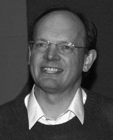
David Lane, 2002.
(Photograph courtesy of CRUK London Research Institute Archives.)
From this, it is clear why p53 loss or mutation is so common in tumours; once it is gone, all hell breaks loose in the genome. If its genome is damaged and cannot be repaired, a cell starts to build up mistakes in its chromosomes every time it replicates. The mistakes are sometimes so catastrophic that the cell dies anyway, but all too often, they result in the loss of other tumour-suppressor genes and the conversion of proto-oncogenes into frank oncogenes, leading to unrestricted growth, and cancer.
p53 is therefore unimportant during a cell’s normal life, yet crucial under battle conditions, explaining how Li-Fraumeni sufferers and their p53-null mouse counterparts can function perfectly well until they encounter situations in which their DNA can be damaged. In spontaneously arising tumours, p53 activity is sometimes lost, just as in Li-Fraumeni syndrome, but it is also frequently mutated into the oncogenic versions that so confused the early researchers in the field. Mutant oncogenic p53 acts as a dominant-negative protein, locking onto any remaining normal p53 and preventing it from working.
Understandably, the p53 pathway has become a prime target for anticancer therapeutics. Although much effort has been expended in trying to design drugs to restore p53 to normality, transcription factors are notoriously hard drug targets, and progress has been slow. However, the prize is so large that the search continues, and it is likely that one or more of the increasingly ingenious strategies will eventually succeed.
And what of David and Lionel? Unsurprisingly, David never went back to immunology and has carried on working on p53 in a career that has turned him into one of the world’s most prominent cancer biologists, and the second most highly cited medical scientist in the United Kingdom in the last decade. He has had an amazing number of very high profile jobs, won a ton of scientific awards, was knighted in 2000, and has been elected to every British society honouring the great and good of science. The bling, however, is not what continues to drive him: “Science is not really a career but much more an obsession—it’s the science that matters above everything. The papers, the grants, the jobs, the prizes are all just surrogates for the science. At times I have been scooped or not followed a line of research that I should have, but in the end, if you honestly do your best, are very persistent, try hard to make a contribution and are generous to others, you will likely succeed” (Lane 2006).
Lionel has had a lower profile. In 1988, he returned to his Cambridge roots, running an ICRF-funded tumour virus lab in the Pathology Department until his retirement in 1995. His subsequent research moved away from SV40 to human papillomaviruses, switching from trying to understand cancer to building on current knowledge to prevent it. It was a worthwhile change, because Lionel’s Cambridge lab was the intellectual birthplace of Human Papilloma Virus vaccines; one of his postdocs, the late Jian Zhou, together with a sabbatical visitor, Ian Frazer, began the work that was the breakthrough for their subsequent development of the Gardasil anti-HPV vaccine, since administered to millions of girls and women to protect them from cervical cancer. Lionel, like David, is a fellow of multiple learned societies, and in 2005, in recognition of his lifetime’s work on DNA tumour viruses, he was awarded the Gabor Medal of the Royal Society. Described by one of his former colleagues as one of the unsung heroes of British biochemistry and molecular biology, it is only appropriate that the last word in this chapter goes to him: “The way science grows is in the cracks between the paving stones. I was a biochemist and David was an immunologist and we thought differently, sometimes at cross-purposes but sometimes synergistically. It was an awfully long time ago, but sometimes it still seems very close.”
Lionel Crawford in retirement.
(Photograph courtesy of Lionel Crawford.)
Further Reading
Lane D, Levine A. 2010. p53 Research: The past thirty years and the next thirty years. Cold Spring Harb Perspect Biol 2: a000893.
Soussi T. 2010. The history of p53. A perfect example of the drawbacks of scientific paradigms. EMBO Rep 11: 822–826.
Two accounts of their research from the horses’ mouths.
Quotation Sources
Mitchison, Avrion. Web of Stories interview. http://www.webofstories.com/play/52547?o=S&srId=397632.
Lane DP, Robbins AK. 1978. An immunochemical investigation of SV40 T antigens. 1. Production properties and specificity of rabbit antibody to purified simian virus 40 large-T antigen. Virology 87: 182–193.
Lane DP, Crawford LV. 1979. T antigen is bound to a host protein in SV40-transformed cells. Nature 278: 261–263.
Lane DP. 1992. p53, guardian of the genome. Nature 358: 15–16.
Lane DP. 2006. Such an obsession. Cancer Biol Ther 5: 120–123.